
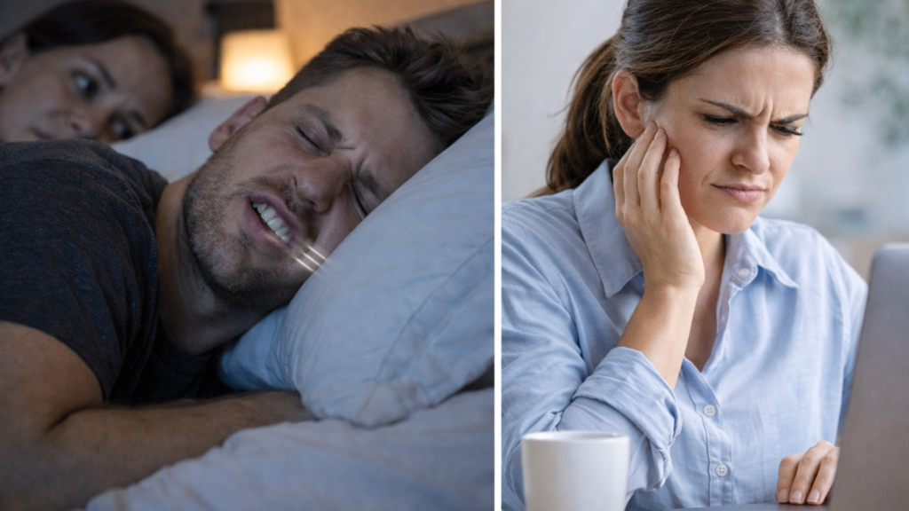
Teeth grinding (bruxism) is a common habit where you clench or grind your teeth, often during sleep, without realizing it. Many people only discover it when they wake up with jaw soreness, headaches, or tooth sensitivity, or when a partner hears grinding sounds at night. While mild grinding may not cause major harm, frequent bruxism can slowly wear down enamel, crack fillings, and overload the jaw joint.
The good news is that bruxism is manageable once you identify the signs and the trigger behind it, stress, sleep quality, bite strain, or lifestyle factors. In this guide, you’ll learn how to spot early symptoms, what kinds of damage to watch for, and when a dentist should check for cracks or TMJ strain. We’ll also break down night guard options, OTC vs custom, soft vs hard, so you can choose a guard that protects your teeth comfortably and fits your needs.
Bruxism Explained: What Teeth Grinding Is and Why It Happens
Bruxism is involuntary clenching or grinding that happens either during sleep or while you’re awake, often without you noticing. Common triggers include stress, poor sleep, caffeine/alcohol, certain medications, and sometimes bite or jaw-muscle strain. Understanding whether yours is sleep bruxism or daytime clenching helps choose the right treatment and night guard.
What is bruxism (sleep vs awake), and how common is it?
Bruxism appears in two distinct forms: sleep bruxism (grinding during sleep) and awake bruxism (daytime clenching).
Sleep bruxism occurs unconsciously during sleep cycles, often intensifying during REM sleep phases. Partners frequently notice the grinding sound before the person realizes they have the condition. Studies indicate approximately 8 to 10% of adults experience sleep bruxism regularly.
Awake bruxism involves conscious or semi-conscious jaw clenching throughout the day, typically during concentration, stress, or physical effort. This form affects roughly 20% of adults at some point, though many cases remain mild. You might clench while driving, working at a computer, or lifting heavy objects without realizing the habit exists.
Why people grind: stress, muscle overactivity, bite factors, lifestyle triggers
Bruxism develops from multiple overlapping factors rather than a single cause.
- Psychological stress ranks as the primary driver for most adults. The jaw muscles carry tension similarly to shoulder or neck muscles, clenching in response to anxiety, work pressure, or emotional strain. Stress-related bruxism often worsens during difficult life periods and improves with stress management.
- Muscle hyperactivity in the jaw plays a role independent of stress. Some individuals have naturally overactive masseter or temporalis muscles that contract more forcefully than necessary during normal function. This biological tendency can persist regardless of emotional state.
- Bite alignment affects grinding patterns in certain cases. Malocclusion (misaligned bite), missing teeth, or poorly fitted dental restorations create uneven contact points. The jaw attempts to “correct” these irregularities through grinding movements, particularly during sleep when conscious control disappears.
- Lifestyle factors amplify bruxism frequency and intensity. Caffeine consumption beyond 2 to 3 cups daily increases muscle tension; alcohol disrupts sleep architecture and triggers more grinding episodes; recreational drugs (especially stimulants) directly increase jaw muscle activity; smoking affects neurotransmitter balance, worsening nighttime grinding.
Bruxism vs TMJ disorder: what’s different and what overlaps
Bruxism and temporomandibular joint (TMJ) disorder represent distinct conditions that frequently coexist and complicate each other.
Bruxism describes the grinding behavior itself, the involuntary muscle activity causing tooth-to-tooth contact. TMJ disorder (temporomandibular disorder or TMD) describes dysfunction in the jaw joint and surrounding structures, causing pain, clicking, limited opening, or locking.
Chronic bruxism can cause TMJ disorder by overloading the joint cartilage, inflaming the joint capsule, and straining the muscles controlling jaw movement. Conversely, existing TMJ dysfunction sometimes triggers grinding as the jaw attempts to find a comfortable resting position.
The overlap creates diagnostic challenges. You might have bruxism without TMJ symptoms (tooth wear with no pain), TMJ disorder without grinding (joint clicking from old injury), or both conditions reinforcing each other (grinding worsens joint inflammation, which increases protective muscle clenching).
Distinguishing the primary problem guides treatment. Grinding-dominant cases respond well to night guards and stress management. TMJ-dominant cases require joint-focused treatments such as physiotherapy, anti-inflammatory protocols, or bite adjustments. Mixed cases need comprehensive approaches addressing both components.
When bruxism is harmless vs when it needs treatment (monitor vs act)
Mild grinding without symptoms or dental damage requires monitoring rather than immediate intervention.
You can safely observe bruxism if you have minimal tooth wear (slight flattening on chewing surfaces that has remained stable for years), no jaw pain or morning stiffness, no headaches attributed to grinding, no damaged fillings or crowns, and a dentist confirms no concerning progression.
Many people grind lightly throughout life without consequences. The teeth possess natural wear resistance, and occasional grinding episodes during stressful weeks rarely cause lasting harm.
Treatment becomes necessary when you notice progressive tooth wear (shortening teeth, exposed dentin, sharp edges developing over months), dental damage (cracked teeth, broken fillings, fractured crowns), persistent jaw symptoms (morning pain, difficulty chewing, clicking that worsens), headaches upon waking or during the day, or sleep disruption affecting daytime function.
The progression rate matters more than a single observation. A dentist comparing wear patterns across 6-month or 12-month intervals identifies accelerating damage requiring intervention versus stable, long-standing wear needing only continued monitoring.
Bruxism and sleep apnea: who should consider screening (key red flags)
Sleep bruxism and obstructive sleep apnea (OSA) share a complex relationship requiring careful screening in specific cases.
Research shows people with sleep apnea grind their teeth at higher rates than the general population. The grinding may serve as an unconscious attempt to reposition the jaw forward and open the airway during breathing interruptions. This protective mechanism becomes problematic when it damages teeth.
You should pursue sleep apnea screening if you experience loud snoring reported by a partner, breathing pauses or gasping during sleep, excessive daytime sleepiness despite adequate sleep hours, morning headaches combined with grinding, or a large neck circumference (over 43 cm in men, over 40 cm in women) plus bruxism.
Treating the apnea often reduces grinding frequency significantly. Continuous positive airway pressure (CPAP) therapy or oral appliances repositioning the jaw eliminate the breathing obstructions, removing the trigger for protective grinding. Addressing only the grinding without treating underlying apnea leaves both conditions inadequately managed.
The connection works both ways. Dentists noticing severe bruxism should ask about sleep quality and snoring. Patients reporting apnea symptoms should inform their dentist, as night guard designs must accommodate airway considerations, some guard types can worsen mild apnea.
Signs and Symptoms: How to Know If You Grind Your Teeth
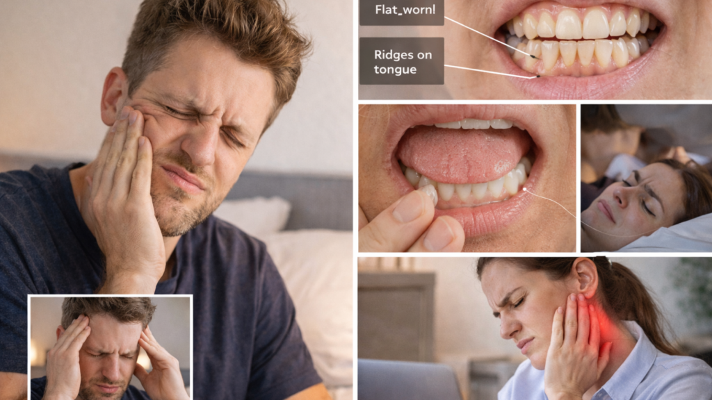
Many people first notice bruxism through morning jaw tightness, headaches, tooth sensitivity, or facial muscle fatigue. Your dentist may spot flat wear marks, tiny cracks, or ridges on the cheeks/tongue that suggest clenching. If you have sharp pain on chewing, swelling, or jaw locking, you should get checked quickly to rule out cracks or TMJ inflammation.
Common symptoms: jaw pain, morning tightness, headaches, neck/shoulder soreness
Bruxism symptoms appear gradually and often worsen overnight, creating a characteristic pattern of morning discomfort.
- Jaw muscle soreness ranks as the most frequent complaint. The masseter muscles (along the jawbone) and temporalis muscles (temples) feel tender, fatigued, or tight upon waking. The discomfort resembles post-exercise muscle soreness from overuse. Symptoms typically improve as the day progresses, only to return the following morning.
- Morning jaw tightness makes opening your mouth fully difficult for the first 30 to 60 minutes after waking. You might struggle to fit three fingers stacked vertically between your front teeth, a quick range-of-motion test. The stiffness gradually releases as you move your jaw through normal daily activities.
- Headaches from bruxism concentrate in specific patterns. Temporal headaches affect the sides of the head where grinding muscles attach. Tension-type headaches span the forehead or back of the head from sustained muscle contraction. These headaches start dull upon waking and either fade or intensify depending on daytime stress levels.
- Neck and shoulder tension extends from jaw muscle strain. The jaw muscles connect anatomically to neck stabilizers and shoulder elevators. Chronic clenching creates referred pain radiating down the neck into the upper shoulders, often mistaken for postural problems or separate muscle strains.
Tooth clues: sensitivity, “worn edges,” chips, cracked fillings, chewing discomfort
Dental evidence often reveals bruxism before you notice symptoms, making regular dental exams essential for early detection.
- Tooth sensitivity from enamel wear increases as grinding wears through protective enamel, exposing underlying dentin. You might experience sharp pain with ctic painful release pattern. Cracks can expose the nerve or propagate into the root, rold drinks, sweet foods, or air hitting the teeth. The sensitivity typically affects multiple teeth rather than a single problem tooth.
- Worn edges appear as flattened chewing surfaces or shortened tooth height. Front teeth develop straight horizontal edges instead of natural rounded contours. Back teeth show cupping (concave wear patterns) on chewing surfaces. Comparing old photos sometimes reveals visible tooth shortening over several years.
- Chips and fracture lines occur along weakened enamel areas. You might notice small pieces breaking off tooth edges or vertical crack lines visible on front teeth when light shines through them. These cracks may not cause immediate pain but indicate structural compromise requiring monitoring.
- Cracked or loose fillings result from grinding forces exceeding the filling material strength. Composite fillings on chewing surfaces fracture or develop gaps at margins. Older amalgam fillings loosen as the surrounding tooth structure wears away. You might feel rough edges with your tongue or notice food packing around previously sealed fillings.
- Chewing discomfort develops when tooth structure becomes thin or nerve irritation increases. Biting on hard foods creates sharp pain or aching that lingers. The discomfort worsens throughout the day as inflammation builds from continued use.
Night-time clues: partner hears grinding, poor sleep, waking with sore jaw
Sleep bruxism often requires external observation for detection, as you remain unconscious during the grinding episodes.
- A partner hearing grinding sounds provides the most direct evidence of sleep bruxism. The noise ranges from faint clicking to loud scraping or gnashing audible across the room. Recording yourself sleeping using a smartphone app can capture the sounds if you sleep alone.
- Poor sleep quality despite adequate sleep hours suggests disrupted sleep architecture from bruxism. You might wake repeatedly without remembering, experience non-restorative sleep leaving you tired, or have difficulty maintaining deep sleep phases. Sleep bruxism intensifies during lighter sleep stages and REM periods.
- Waking with a clenched jaw or sore muscles indicates grinding occurred overnight. You might notice jaw tightness immediately upon waking or catch yourself grinding during brief nighttime awakenings. Some people wake from particularly intense grinding episodes.
Unusual tongue or cheek marks appear from grinding pressure. Scalloped tongue edges (indentations matching tooth patterns) or ridged cheek tissue along the bite line develop from sustained pressure against teeth during clenching. These tissue changes persist throughout the day.
Quick self-check at home (and what it can’t confirm)
A basic home assessment helps identify potential bruxism, though professional evaluation remains necessary for definitive diagnosis.
- Check jaw muscle tenderness by pressing fingertips firmly along the jawbone angle (masseter muscles) and temples (temporalis muscles). Sore or tight muscles compared to surrounding areas suggest overuse from grinding. Compare both sides, as grinding often affects one side more severely.
- Examine teeth in a mirror for wear patterns. Look for flattened chewing surfaces on back teeth, shortened or squared-off front teeth, or shiny worn spots on enamel. Run your tongue along tooth edges feeling for sharp corners or rough areas indicating chips.
- Test jaw opening range by stacking three fingers vertically and attempting to fit them between your front teeth. Difficulty accommodating three fingers indicates restricted opening from muscle tightness or joint limitation.
- Monitor morning symptoms by noting jaw soreness, headaches, or facial fatigue upon waking for 7 to 10 days. Patterns emerging most mornings suggest nighttime bruxism.
Home checks cannot confirm the diagnosis definitively, determine grinding severity, identify underlying causes (stress vs sleep disorder vs bite problem), assess dental damage extent, or rule out other conditions mimicking bruxism. Professional evaluation provides these critical assessments guiding appropriate treatment.
Warning signs that need urgent dental evaluation (crack pain, swelling, locking jaw)
Certain symptoms indicate severe bruxism complications requiring prompt professional assessment rather than waiting for routine appointments.
- Sudden sharp pain on biting suggests a cracked or broken tooth. The pain intensifies with chewing and release, creating a characteristic painful release pattern. Cracks can expose the nerve or propagate into the root, risking tooth loss without rapid treatment.
- Swelling in the jaw, face, or gums indicates possible infection or abscess formation. Grinding-related cracks provide bacterial entry points. Swelling accompanied by fever, difficulty swallowing, or spreading redness requires same-day evaluation.
- Jaw locking in an open or closed position represents acute TMJ dysfunction. You cannot move the jaw voluntarily, or movement produces severe pain. Locking episodes suggest displaced disc or severe joint inflammation requiring specialized management.
- Tooth mobility or looseness develops from extreme grinding forces damaging the periodontal ligament anchoring teeth to bone. Movement visible when pushing the tooth with your finger or looseness felt when chewing needs immediate assessment to prevent tooth loss.
- Numbness or tingling in the face, lips, or tongue can indicate nerve compression from severe muscle swelling or refer from spinal issues worsened by jaw posturing. Neurological symptoms require ruling out serious conditions.
These warning signs merit contacting a dentist within 24 to 48 hours rather than delaying until scheduled checkups. Early intervention for complications prevents extensive damage and improves treatment outcomes.
Damage Bruxism Can Cause: Teeth, Gums, Jaw, and Restorations
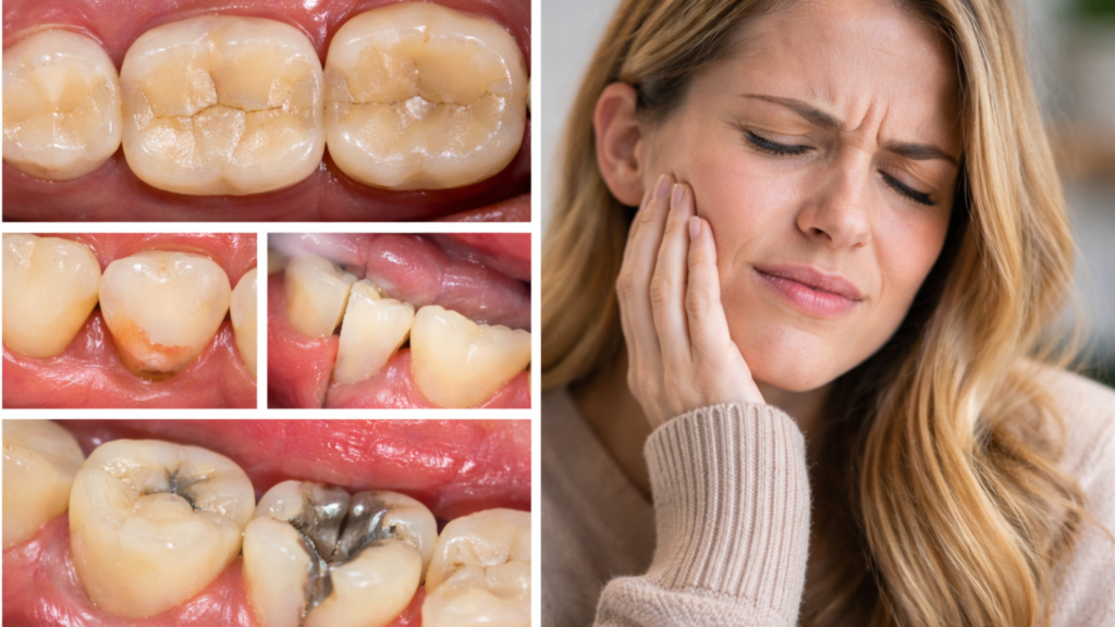
Frequent grinding can wear enamel, chip teeth, crack fillings, and shorten the lifespan of crowns or veneers. It may also contribute to gum recession and “notches” near the gumline from repeated overload. Over time, jaw muscles and the TMJ can become irritated, leading to clicking, pain near the ear, or limited opening.
Tooth damage: enamel wear, fractures, cracked teeth, loose fillings, crown failures
Tooth structure bears the direct impact of grinding forces, accumulating damage over months to years of untreated bruxism.
- Enamel wear progresses from outer protective layers into softer underlying dentin. Teeth appear shorter, flatter, or more yellowed as dentin (naturally yellow) shows through thinning enamel. The wear reduces tooth height by 1 to 3 millimeters in moderate cases and over 5 millimeters in severe long-term grinding, altering facial appearance and bite relationships.
Fractures develop in predictable patterns based on force direction. Vertical cracks run from the chewing surface toward the root, often starting unnoticed and propagating deeper over time. Horizontal fractures break off tooth sections completely. Front teeth chip at edges where enamel is thinnest.
- Cracked teeth create diagnostic challenges because cracks may not show on X-rays until advanced. Symptoms include pain on biting that disappears upon release, sensitivity to temperature changes, or intermittent sharp pain without obvious cause. Untreated cracks extend below the gumline, often requiring extraction rather than restoration.
- Loose fillings occur because grinding forces exceed filling material adhesion strength. Composite fillings separate at margins, creating gaps allowing bacterial invasion and decay. Large fillings weaken remaining tooth structure, making fractures more likely under grinding stress.
- Crown failures happen through several mechanisms. Porcelain crowns chip or fracture on chewing surfaces. Crown cement breaks down from repeated stress, causing looseness. The underlying tooth structure fractures beneath the crown, requiring removal and retreatment or extraction.
The cumulative damage increases exponentially with time. What starts as minor enamel wear progresses to fractures requiring crowns, then to crown failures needing extractions and implants, each stage more complex and expensive than early intervention with protective appliances.
Gum and bone strain: recession, abfraction lesions, bite overload
Grinding forces transmit through teeth into supporting gums and bone, causing tissue damage beyond the teeth themselves.
- Gum recession from bite overload accelerates from excessive biting forces pushing teeth slightly out of normal position. The gum tissue pulls away from tooth surfaces, exposing root surfaces. Recession typically affects the side of teeth receiving maximum force, creating asymmetric patterns distinguishing it from recession caused by aggressive brushing.
- Abfraction lesions appear as V-shaped notches near the gumline on tooth surfaces. Grinding forces flex teeth at the narrow neck region, causing enamel to fracture away microscopically over time. These lesions differ from cavities because they occur in plaque-free areas and have sharp, angular shapes rather than the rounded contours of decay.
- Bone loss around tooth roots develops from bite overload. Excessive force exceeds the periodontal ligament’s adaptive capacity, causing inflammation and gradual bone resorption. X-rays show widened periodontal ligament space around affected teeth or decreased bone height. Bone loss destabilizes teeth, potentially leading to looseness or tooth loss in extreme cases.
The tooth-bone interface (periodontal ligament) normally absorbs and distributes biting forces. Bruxism generates forces 2 to 10 times normal chewing strength, overwhelming this system. Repeated overload creates chronic inflammation, triggering the body’s bone-remodeling processes in destructive rather than protective patterns.
Combination damage proves most destructive. Recession exposes sensitive root surfaces; abfraction lesions weaken structural integrity; bone loss reduces tooth support, together creating conditions for tooth loss even without cavities or gum disease.
Jaw joint and muscles: TMJ pain, clicking, limited opening, facial fatigue
The temporomandibular joint and surrounding muscles endure sustained stress from grinding, developing pain and dysfunction over time.
- TMJ pain manifests as aching or sharp pain directly in front of the ear where the jaw hinge articulates. Pain worsens with jaw movement (chewing, talking, yawning) and improves with rest. Some people feel a deep, dull ache versus sharp catching pain indicating different joint structure involvement.
- Clicking or popping sounds during jaw opening occur when the articular disc (cartilage cushion inside the joint) slips out of proper position. The click represents the disc relocating as the jaw moves. Painless clicking alone does not require treatment, but clicking accompanied by pain or progression to locking indicates advancing joint damage.
- Limited jaw opening develops from muscle guarding (protective spasm preventing movement) or true joint restriction (internal derangement blocking motion). You might open only 25 to 35 millimeters between front teeth instead of the normal 40 to 50 millimeters. Restriction worsens morning symptoms after nighttime grinding.
- Facial muscle fatigue creates a tired, heavy feeling in the jaw and cheeks. Muscles feel overworked from sustained contraction, similar to leg fatigue after excessive exercise. The fatigue interferes with normal activities such as eating, speaking, or maintaining comfortable jaw posture.
Muscle trigger points form as tight, painful knots in overworked jaw muscles. Pressing these points reproduces familiar pain patterns or refers pain to the temples, neck, or behind the eyes. Trigger points perpetuate the pain cycle even when grinding reduces.
Chronic muscle tension alters jaw movement patterns. You might develop asymmetric opening (jaw deviates to one side) or difficulty achieving a comfortable bite position. These altered patterns reinforce abnormal muscle firing, sustaining the problem beyond the original grinding trigger.
Headache and ear symptoms: temples pain, ear fullness, ringing (why it happens)
Bruxism creates referred pain patterns affecting the head and ears through anatomical connections and muscle tension pathways.
- Temple headaches occur because the temporalis muscle (primary grinding muscle) attaches across the temple area. Sustained clenching creates muscle tension that refers pain throughout its attachment zone. The pain feels like pressure or aching rather than sharp or throbbing, distinguishing it from migraines.
- Ear fullness or pressure happens through two mechanisms. Firstly, muscles of mastication (chewing muscles) attach near the Eustachian tube opening, and muscle tension can affect tube function, creating a blocked sensation. Secondly, the TMJ sits immediately in front of the ear canal, and joint inflammation or swelling causes fullness sensations mimicking ear problems.
- Ringing in the ears (tinnitus) connects to TMJ dysfunction through poorly understood pathways. Theories include nerve pathway overlap between jaw and ear structures, altered blood flow from muscle tension affecting inner ear function, or changes in middle ear muscle tone from grinding-related neural activation. The ringing typically fluctuates with jaw symptoms, worsening during intense grinding periods.
Pain behind the eyes develops from referred tension in the temporalis muscle’s anterior fibers. These muscle sections attach near the eye socket, creating pain patterns easily confused with sinus problems or eye strain. The pain lacks the pulsing quality of migraines and improves with jaw rest or muscle massage.
These symptoms drive many patients to ear, nose, and throat specialists before discovering the jaw connection. Ear exams show normal structures; sinus X-rays reveal no pathology, finally leading to dental evaluation revealing bruxism as the hidden cause. Understanding these referred pain patterns prevents unnecessary testing and targets treatment effectively.
Diagnosis at a Dental Clinic in Kathmandu: What to Expect
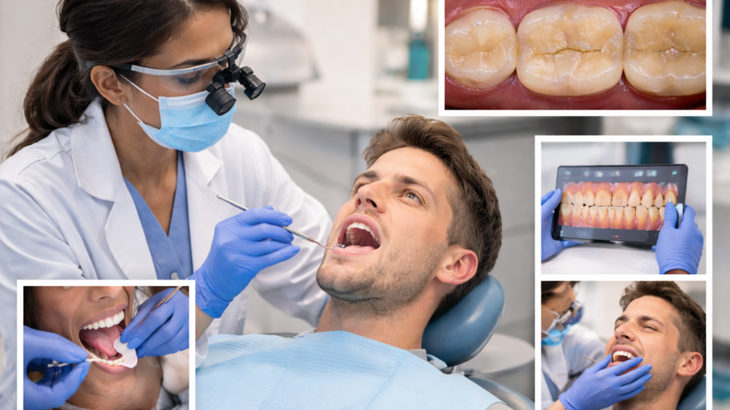
A dental exam for bruxism usually includes checking tooth wear patterns, bite balance, muscle tenderness, and any signs of cracks. X-rays may be recommended if there’s sensitivity, chewing pain, or older fillings to assess hidden damage. If symptoms suggest sleep issues like snoring or daytime sleepiness, your dentist may advise screening for sleep-disordered breathing.
The dental exam checklist: wear pattern mapping, bite check, muscle tenderness
A thorough bruxism assessment at BrightSmile Dental Clinic follows a systematic protocol identifying grinding signs and underlying causes.
Wear pattern mapping documents tooth surface damage systematically. The dentist examines each tooth noting flattened cusps, exposed dentin (yellowish areas), sharp edges, or polished facets (shiny spots indicating grinding). Patterns reveal grinding direction and intensity, generalized wear suggests sleep bruxism affecting all teeth, while localized wear indicates specific bite interferences.
The dentist compares wear patterns against age-expected wear. Moderate wear at age 60 differs from identical wear at age 30, where it signals aggressive bruxism requiring intervention. Documentation establishes a baseline for monitoring progression over subsequent visits.
Bite assessment checks how upper and lower teeth contact in various jaw positions. The dentist evaluates centric occlusion (maximum intercuspation), looks for premature contacts (single teeth hitting before others), and tests lateral movements for guidance patterns. Bite irregularities concentrate forces on specific teeth, driving localized grinding patterns.
Muscle palpation identifies tender or hypertrophic muscles. The dentist presses masseter muscles along the jawbone, temporalis muscles at the temples, medial pterygoid muscles inside the mouth, and neck muscles checking for pain, spasm, or excessive muscle development. Asymmetric muscle tenderness reveals unilateral grinding patterns or compensatory muscle behaviors.
Range of motion testing measures maximum opening (normal 40 to 50 mm), lateral movements, and protrusive movement. Restricted or painful movement indicates muscle or joint involvement requiring specific treatment approaches beyond simple tooth protection.
When X-rays help: hidden cracks, failed fillings, deep tooth damage
Radiographic imaging reveals bruxism damage invisible during clinical examination, guiding treatment planning and urgency assessment.
Periapical X-rays (detailed images of individual teeth) show vertical root fractures, widened periodontal ligament space indicating bite trauma, bone loss around tooth roots, and internal resorption from chronic inflammation. These findings change treatment recommendations significantly, for instance, a cracked root requires extraction rather than a simple filling.
Bitewing X-rays detect decay under failing fillings, cracked filling margins, and bone level changes. Grinding loosens old fillings, creating gaps allowing bacterial invasion and secondary decay invisible on tooth surfaces. Early detection prevents small problems from becoming root canals.
Panoramic X-rays provide an overview showing jaw joint condition, overall bone levels, and relationship of tooth roots to surrounding structures. Some practitioners request these for comprehensive TMJ assessment when joint symptoms accompany grinding.
Not every bruxism case requires X-rays immediately. The dentist orders imaging when you have symptoms suggesting hidden damage (intermittent pain without obvious cause, previous dental trauma history, old or large fillings in grinding zones), visible cracks needing depth assessment, or monitoring progression in severe cases. Radiation exposure follows ALARA principles (as low as reasonably achievable).
Digital radiography at modern Kathmandu clinics like BrightSmile reduces radiation exposure by 50 to 80% compared to traditional film X-rays while providing instant, enlarged images improving diagnostic accuracy.
Identifying the “main driver”: stress clenching vs sleep bruxism vs bite issues
Effective treatment requires determining which factor predominantly drives your grinding, stress, sleep disorders, or dental causes.
Stress-related daytime clenching shows specific patterns. You might report awareness of clenching during work, driving, or concentration. The clenching increases during stressful periods and decreases during vacations. Muscle tenderness concentrates in masseter muscles. Tooth wear appears more on back teeth bearing maximum clenching force. Daytime bruxism responds well to awareness training and stress management.
Sleep bruxism presents differently. A partner reports nighttime grinding sounds. You have no awareness of the habit. Morning symptoms (jaw soreness, headaches) dominate, improving through the day. Wear patterns affect front and back teeth equally from unconscious jaw movements. Sleep bruxism requires protective night guards since conscious control does not exist during sleep.
Bite-driven grinding shows identifiable dental triggers. Recent dental work preceded symptom onset. You notice specific teeth hitting prematurely when biting. Grinding attempts to “fix” the interference through wear. Wear concentrates on teeth involved in the premature contact. Bite-driven cases respond to occlusal adjustment or bite equilibration addressing the structural problem.
Many patients show mixed patterns requiring combined approaches. You might clench during the day from stress and grind at night from sleep disruption, needing both behavior modification and night guard protection. The dentist weighs the dominant factor, initiating treatment there while planning sequential addition of other strategies.
Identifying the main driver prevents ineffective treatment. Providing a night guard to someone with primarily daytime stress clenching addresses only partial of the problem. Adjusting the bite for someone with pure sleep bruxism from anxiety misses the target entirely.
When sleep problems are suspected: snoring, daytime sleepiness, referral pathway
Bruxism accompanied by sleep disorder symptoms requires coordinated assessment beyond standard dental care.
Red flag symptoms prompting sleep evaluation include loud snoring reported by a partner, witnessed breathing pauses or gasping during sleep, excessive daytime sleepiness despite adequate sleep hours (falling asleep during meetings, while driving), morning headaches plus bruxism, or obesity plus grinding. These patterns suggest obstructive sleep apnea driving protective grinding.
The referral pathway typically starts with the dentist documenting sleep symptoms and recommending sleep medicine consultation. Sleep specialists conduct comprehensive assessment including sleep history, Epworth Sleepiness Scale, STOPBANG questionnaire, and possibly home sleep testing or formal polysomnography (sleep study).
Sleep studies record brain waves, oxygen levels, breathing patterns, heart rate, and muscle activity through the night. The results quantify apnea severity through AHI (apnea-hypopnea index), correlate breathing events with grinding episodes, and guide treatment recommendations.
Treatment for confirmed sleep apnea often reduces bruxism significantly. CPAP therapy maintains open airways, eliminating the physiological trigger for protective grinding. Oral appliances repositioning the jaw forward improve airway patency in mild to moderate cases while simultaneously reducing grinding.
The dentist and sleep specialist collaborate on device selection. Some oral appliances serve dual purposes, advancing the jaw to open airways while protecting teeth from grinding forces. Others require separate management of each condition.
Patients with sleep apnea should inform the dentist before receiving standard night guards. Bulky guards can worsen mild apnea by displacing the tongue or mandible. Guards for apnea patients need specific design modifications or coordination with CPAP use.
Night Guard Options: Types, Fit, Comfort, and Choosing the Right One
Night guards mainly protect your teeth from damage and reduce overload on jaw muscles, but they don’t always “stop” the grinding habit. Custom guards made by a dentist typically fit better, last longer, and are safer for your bite than many OTC boil-and-bite guards. The best choice depends on how hard you clench, whether you have TMJ pain, and how sensitive you are to bulky appliances.
Soft vs hard vs dual-laminate: which guard suits which grinding pattern
Night guard material selection significantly impacts protection effectiveness, comfort, and grinding behavior modification.
- Soft guards use flexible silicone or thermoplastic materials providing cushioning comfort. The soft texture feels gentle against teeth and gums. However, soft guards can encourage chewing behavior in some people, actually increasing grinding frequency or intensity. The material compresses under force rather than redirecting it, offering less structural protection for severe grinders. Soft guards suit mild, occasional grinders who need comfort prioritization or initial guard users testing tolerance.
- Hard acrylic guards use rigid plastic materials that do not compress under biting forces. The hard surface prevents teeth from achieving full grinding contact, reducing force transmission through tooth structure. Hard guards discourage grinding by providing an unpleasant sensation when attempting to grind, sometimes retraining muscle patterns over time. Hard guards suit moderate to severe grinders, people with significant tooth wear or fractures, and those committed to long-term protection.
- Dual-laminate guards combine a soft inner layer contacting teeth with a hard outer layer receiving opposing teeth. The design balances comfort and protection, soft interior reduces irritation while hard exterior provides structural strength. Dual-laminate guards suit people who find hard guards too uncomfortable but need better protection than soft materials provide, moderate grinders, and patients with sensitive teeth or gums.
Material thickness matters equally. Thin guards (2 to 3 mm) offer minimal bulk but less protection and shorter lifespan. Thick guards (4 to 5 mm) maximize protection and durability but increase initial discomfort and adjustment period. The dentist selects thickness based on grinding severity and available space.
OTC boil-and-bite vs dentist-made custom: safety, comfort, durability, protection
Over-the-counter (OTC) and custom-fabricated guards differ dramatically in fit, effectiveness, and long-term value.
- OTC boil-and-bite guards soften in hot water, allowing you to bite down and create a generalized impression. The guards cost NPR 800 to 2,500, provide same-day use, and require no dental visit. However, the fit remains imprecise, bulky, loose in areas, or uneven thickness. Poor fit creates gagging, difficulty breathing, tooth movement over time, or jaw discomfort from altered bite position. OTC guards suit very short-term use during high-stress periods, travel emergencies, or trialing guard tolerance before investing in custom options.
- Custom night guards require impressions or digital scans of your teeth, fabrication in a dental laboratory to your exact anatomy, and professional fitting with adjustments. The cost ranges from NPR 8,000 to 16,000 in Kathmandu clinics like BrightSmile. The investment provides precise fit minimizing bulk, stable retention preventing dislodgement during sleep, even thickness distributing forces properly, comfortable margins preventing tissue irritation, and controlled bite relationships avoiding jaw position problems.
The fit precision makes the difference. Custom guards contact all teeth evenly, stabilizing the jaw position and distributing grinding forces across the entire arch rather than concentrating stress on a few teeth. Proper occlusal (bite) surface design on custom guards guides jaw movements safely.
Durability varies significantly. OTC guards wear through or deform within 3 to 6 months of nightly use. Custom hard acrylic guards last 2 to 5 years with proper care. The longer lifespan offsets higher initial cost.
Safety concerns with OTC guards include potential tooth movement from uneven forces, TMJ problems from improper bite positioning, and inadequate material thickness allowing breakthrough grinding damage. Custom guards designed by dental professionals avoid these risks through controlled fabrication.
Full-coverage vs partial guards: why full coverage is usually safer long-term
The extent of tooth coverage affects protection effectiveness, bite stability, and potential side effects.
Full-coverage guards extend over all upper (or lower) teeth from molars to front teeth. The continuous coverage distributes forces evenly across the entire arch. Full coverage stabilizes the jaw position, prevents individual tooth movement, protects all teeth equally, and creates a stable platform for jaw function.
Partial guards cover only front teeth or a section of the arch. The reduced material minimizes bulk and improves initial comfort. However, partial coverage creates several problems. Uncovered teeth receive concentrated grinding forces, often causing damage to unprotected areas. The bite rests only on front teeth, overloading those structures. Posterior teeth can erupt or shift over time from lack of contact (supereruption), creating permanent bite changes.
Anterior deprogrammers (NTI devices) cover only front teeth, attempting to reduce muscle activity through neurological mechanisms. These devices carry risks including posterior tooth supereruption within weeks to months, bite changes difficult to reverse, and TMJ problems from altered jaw positioning. Anterior devices suit only very short-term use (days to weeks) under close supervision.
Full coverage suits nightly long-term use, moderate to severe grinding, people with existing TMJ issues, and anyone with dental restorations (crowns, implants) requiring comprehensive protection. Partial guards might work for very mild grinding, short-term evaluation periods, or specific situations where full coverage proves impossible due to strong gag reflex.
The consensus among dental specialists favors full-coverage guards for sustained bruxism management. Initial discomfort from slightly larger appliances represents a worthwhile trade-off for comprehensive protection and bite stability.
What a night guard can and can’t do (protect teeth vs “stop grinding”)
Understanding night guard capabilities and limitations sets realistic expectations for treatment outcomes.
Night guards can protect teeth from wear, fractures, and damage by absorbing grinding forces; reduce muscle soreness and morning stiffness in many users by providing a stable jaw position; prevent further damage to existing dental work (crowns, fillings, implants); reduce headache frequency and intensity in grinding-related cases; and improve sleep quality by reducing arousal episodes from grinding intensity.
Night guards cannot stop the grinding behavior itself, you will continue grinding, but on the guard instead of teeth; cure underlying causes (stress, sleep apnea, anxiety); correct bite problems or misaligned teeth; treat TMJ disorders without additional therapies; or provide permanent solutions, removing the guard returns you to risk status.
The guard functions as a protective device similar to a safety helmet, it protects against injury from the activity without preventing the activity itself. Some users experience reduced grinding frequency over months of guard use through neural adaptation, but this secondary benefit occurs inconsistently.
Realistic expectations improve satisfaction and treatment adherence. You should expect tooth protection and symptom reduction, not grinding cessation. Combine the guard with addressing underlying triggers (stress management, sleep disorder treatment, bite adjustment if needed) for optimal long-term results.
How a custom night guard is made: scan/impression → lab → fitting → adjustments
Custom night guard fabrication at BrightSmile Dental Clinic follows a precise multi-step process ensuring optimal fit and function.
The initial appointment involves examination confirming bruxism, discussing guard types and materials, and capturing your dental anatomy. The dentist takes impressions using either traditional putty material (which you bite into until set) or digital intraoral scanning (a camera wand mapping tooth surfaces). Digital scans provide greater comfort and precision. The dentist also records your bite relationship using wax or digital registration.
Laboratory fabrication takes 5 to 7 days typically. Skilled technicians pour models from impressions, design the guard accounting for your specific grinding pattern and bite, fabricate the guard through vacuum forming or milling, and trim and polish for smooth comfortable margins.
The fitting appointment occurs once the guard returns from the lab. The dentist places the guard, checks retention and stability, evaluates comfort and any pressure points, adjusts the bite surface for even contact, trims any sharp edges or excessive bulk, and demonstrates insertion, removal, and care. You wear the guard for a few minutes to identify any discomfort spots requiring adjustment.
Most patients need one or two additional adjustment visits. You return after 1 to 2 weeks of nightly use reporting any issues. The dentist addresses pressure points causing soreness, areas causing gagging, uneven bite feel, or looseness. Small adjustments dramatically improve comfort and compliance.
Total timeline from initial visit to final adjusted guard spans 2 to 3 weeks. The investment in proper fabrication and fitting pays through years of effective protection.
Troubleshooting: if the guard hurts, causes gagging, smells, cracks, or feels tight
Common night guard problems have straightforward solutions when addressed promptly with your dentist.
Guard pain typically comes from pressure points or uneven bite contact. Localized sore spots indicate overly thick areas pressing into gum tissue, the dentist trims these areas. Generalized soreness suggests clenching harder on the guard initially (usually resolves within one week as you adapt) or uneven occlusion requiring bite adjustment.
Gagging occurs from guards extending too far toward the throat or being overly thick. The dentist shortens the posterior extension by several millimeters, thins the palatal area (roof portion), or switches to a lower guard if you have a sensitive gag reflex. Most gag reflexes improve with gradual adaptation, starting with short wear periods (30 minutes before bed) and increasing nightly.
Odor and discoloration develop from bacterial buildup or inadequate cleaning. Clean the guard daily using a soft toothbrush and mild soap (not toothpaste, which is too abrasive). Soak weekly in denture cleaner or diluted vinegar solution (1 part vinegar to 3 parts water for 15 to 20 minutes). Store dry in a ventilated case, moisture encourages bacterial growth.
Cracks or holes indicate the guard is too thin for your grinding intensity or has reached end of lifespan. Severe grinders wear through guards faster, sometimes within 12 to 18 months. Replace cracked guards immediately, they provide inadequate protection and sharp edges can injure soft tissues.
Tightness or difficulty removing the guard suggests minor tooth movement (normal micro-movement happens even in adults) or swollen gums from inadequate cleaning. The dentist can adjust retention or check for gum inflammation requiring improved oral hygiene.
Persistent problems after adjustments may indicate poor initial design. Remaking the guard solves issues better than excessive adjustments in some cases.
Night guard care: cleaning, storage, lifespan, and when to replace
Proper maintenance extends guard lifespan and prevents oral health complications from contaminated appliances.
Daily cleaning after each use prevents bacterial buildup. Rinse the guard under cool water immediately after removal. Brush all surfaces using a soft toothbrush and mild liquid soap or dish detergent, avoid toothpaste because abrasive particles scratch the guard surface, creating bacterial retention sites. Rinse thoroughly and shake off excess water.
Weekly deep cleaning removes deeper biofilm. Soak the guard in denture cleaning solution, diluted white vinegar (1:3 with water), or specialized retainer cleaner for 15 to 20 minutes. Scrub again and rinse thoroughly. Avoid hot water, which can warp the material.
Proper storage maintains guard integrity. Store the guard completely dry in a ventilated case allowing airflow. Never leave it wrapped in tissue or closed wet containers, moisture encourages bacterial and fungal growth. Keep the guard away from heat sources (car dashboards, windowsills, heaters) preventing warping.
Expected lifespan varies by material and grinding severity. Soft guards last 6 to 12 months with nightly use. Hard acrylic guards last 2 to 5 years. Dual-laminate guards last 18 months to 3 years. Severe grinders replace guards more frequently than mild grinders.
Replace your guard when you notice visible wear-through holes or thin spots, cracks or fractures anywhere, significant rough areas or sharp edges, loosening fit that adjustment cannot resolve, or persistent odor despite thorough cleaning. Continuing to use worn-out guards reduces protection effectiveness and increases bacterial contamination risk.
Bring your guard to routine dental checkups. The dentist inspects for damage, assesses wear patterns adjusting treatment if needed, and professionally cleans the guard removing buildup you cannot eliminate at home.
Treatment Beyond Guards: Long-Term Relief, Kids, and Prevention Plan
Long-term control often combines a night guard with trigger management—stress reduction, better sleep habits, and correcting any dental factors that increase strain. Jaw stretches, heat therapy, and physiotherapy can help muscle-related pain, and severe cases may need additional options like Botox under professional guidance. Regular follow-ups help adjust the guard, monitor wear, and prevent cracks before they become emergencies.
Daytime clenching fixes: awareness, habit reversal, jaw relaxation drills
Addressing daytime clenching requires conscious behavior modification since you control jaw position while awake.
Awareness training forms the foundation. You cannot change a habit you do not notice. Set hourly phone reminders labeled “Check jaw” prompting you to assess whether you are clenching. Notice clenching triggers, computer work, driving, stress, concentration, phone conversations. Keep a clenching diary noting when, where, and during what activities you clench most.
The proper rest position keeps teeth apart with lips closed and tongue resting against the palate behind front teeth. This posture relaxes jaw muscles while maintaining facial composure. Practice finding this position repeatedly throughout the day until it becomes habitual.
Habit reversal techniques interrupt the clenching pattern. When you notice clenching, immediately open your mouth wide (gentle yawn), move the jaw side to side, and relax into proper rest position. The interruption breaks the automatic behavior pattern. Repeat consistently whenever you catch yourself clenching.
Jaw relaxation drills reduce baseline muscle tension. Practice gentle jaw stretches (open wide, move side to side, protrude forward) several times daily. Apply warm compresses to jaw muscles for 10 to 15 minutes to promote relaxation. Massage tender points in masseter and temporalis muscles using small circular motions.
Progressive muscle relaxation involves deliberately tensing and relaxing jaw muscles to increase body awareness. Clench lightly for 5 seconds, then release completely for 10 seconds, noticing the difference. This training improves your ability to detect and release unwanted tension.
Address the root stress through stress management techniques beyond jaw-focused interventions. Exercise regularly to reduce systemic stress, practice meditation or deep breathing, ensure adequate sleep, and consider counseling for persistent anxiety or stress-related issues.
Sleep and lifestyle plan: caffeine/alcohol timing, stress reset, better sleep hygiene
Lifestyle modifications reduce grinding triggers and improve sleep quality, complementing physical protective measures.
Caffeine management requires strategic timing rather than complete elimination. Limit total daily caffeine to 200 to 300 milligrams (approximately 2 to 3 cups of coffee). Avoid caffeine after 2 PM, giving your system 8+ hours to metabolize before sleep. The stimulant effect increases muscle tension and disrupts sleep architecture, worsening nighttime grinding.
Alcohol impacts sleep despite its sedating initial effect. Alcohol fragments sleep, increases REM rebound (when most grinding occurs), and reduces sleep quality. Limit alcohol to 1 to 2 drinks maximum and finish drinking 3 to 4 hours before bed. The brief relaxation does not compensate for disrupted sleep architecture.
Stress reset practices improve parasympathetic nervous system activation. Dedicate 15 to 30 minutes before bed to relaxing activities, reading, gentle stretching, warm bath, or meditation. Create a mental transition from day stress to sleep preparation. The intentional wind-down reduces muscle tension carried into sleep.
Sleep hygiene optimization creates conditions for restorative sleep. Maintain consistent sleep and wake times even on weekends to stabilize circadian rhythm. Keep the bedroom cool (18 to 20°C), dark, and quiet. Remove screens (phones, tablets, TVs) 1 hour before bed, blue light suppresses melatonin production. Reserve the bed for sleep and intimacy only, not work or stressful activities.
Exercise timing affects sleep quality. Regular exercise reduces stress and improves sleep, but vigorous activity within 3 hours of bedtime increases alertness and body temperature, delaying sleep onset. Schedule intense workouts for morning or early afternoon.
Sleep position influences jaw position and grinding in some people. Sleeping on your back reduces jaw pressure compared to side or stomach sleeping. Experiment with different positions and pillow heights to find optimal jaw alignment during sleep.
Pain relief and recovery: heat/ice, stretches, physiotherapy, when meds are used
Acute symptom management provides relief while addressing underlying causes through long-term interventions.
Heat therapy relaxes tight muscles and improves blood flow. Apply moist heat (warm towel, heating pad on low setting, warm compress) to jaw muscles for 15 to 20 minutes. The warmth reduces muscle spasm and relieves soreness. Use heat for chronic, ongoing muscle tightness.
Ice therapy reduces acute inflammation and swelling. Apply ice packs (wrapped in cloth to prevent skin damage) for 10 to 15 minutes during acute pain episodes or immediately after excessive clenching periods. Ice constricts blood vessels, reducing inflammation and numbing pain.
Alternating heat and ice can be effective for some patients. Start with ice for 10 minutes to reduce inflammation, follow with heat for 15 minutes to relax muscles, and repeat the cycle 2 to 3 times. The contrast therapy improves circulation and addresses both inflammation and muscle tension.
Jaw stretches maintain mobility and reduce stiffness. Practice gentle opening (open as wide as comfortable, hold 5 seconds, repeat 5 times), lateral movement (move jaw to each side, hold, repeat), and forward protrusion (stick jaw forward gently, hold, return). Perform stretches 2 to 3 times daily, especially after waking.
Physiotherapy or physical therapy addresses chronic muscle dysfunction and TMJ problems. Trained therapists provide manual therapy (joint mobilization, soft tissue massage), teach specific exercises targeting weak or imbalanced muscles, use modalities (ultrasound, electrical stimulation) for pain relief, and educate on posture and ergonomics reducing jaw strain.
Medications serve supportive roles in bruxism management. Over-the-counter NSAIDs (ibuprofen 400 mg every 6 to 8 hours, or naproxen 220 mg every 8 to 12 hours) reduce inflammation and pain during acute episodes, use short-term (3 to 7 days) to avoid side effects. Muscle relaxants (prescribed by physicians) occasionally help severe muscle spasm, typically used for brief periods (1 to 2 weeks) during acute flare-ups.
Medications do not address bruxism causes and carry side effects with prolonged use. Use them as temporary aids while implementing behavior changes, stress management, and protective devices for lasting improvement.
Advanced options (brief): Botox for severe clenching + when it’s considered
Botulinum toxin (Botox) injections represent an advanced intervention for severe, treatment-resistant bruxism.
Botox works by temporarily weakening the masseter and temporalis muscles through blocking nerve signals causing muscle contraction. The injections reduce clenching force intensity without eliminating all jaw function. Chewing and speaking continue normally with somewhat reduced power.
The procedure involves multiple small injections into the bulk of the masseter muscles (along the jawbone) and sometimes temporalis muscles (temples). Effects begin within 3 to 7 days, peak at 2 to 4 weeks, and last approximately 3 to 6 months. Repeat injections maintain benefits.
Botox suits severe grinders with significant tooth damage despite night guard use, chronic jaw pain unresponsive to conservative treatments, masseter hypertrophy (visibly enlarged jaw muscles), or headaches from muscle tension not controlled by other methods. The treatment works best as part of comprehensive management, not as a sole solution.
Costs in international markets range from NPR 30,000 to 80,000 per session (prices vary by provider and units required). The expense and temporary nature make Botox appropriate only after conservative measures fail.
Potential side effects include temporary difficulty chewing very tough foods, slight asymmetry if injection placement is uneven, bruising at injection sites, or headache. Serious complications are rare when performed by trained practitioners.
Botox availability in Kathmandu is expanding through specialized dental and dermatology practices. Discuss the option with your dentist if conventional treatments provide insufficient relief.
Teeth grinding in children: causes, reassurance, and when parents should worry
Childhood bruxism differs significantly from adult grinding in causes, implications, and treatment approaches.
Grinding in children occurs commonly, affecting 20 to 30% of children at some point during development. Causes in children include teething discomfort (ages 6 months to 3 years), jaw growth and tooth development, airway issues (enlarged tonsils or adenoids obstructing breathing), stress or anxiety (school pressure, family changes), and sleep disorders.
Most childhood grinding resolves spontaneously without intervention. The habit typically peaks during tooth eruption periods and disappears once permanent teeth fully emerge (around ages 6 to 7, and again around 10 to 13). The developing jaw adapts to changing bite relationships through grinding movements.
Parents should monitor rather than immediately treat when grinding is mild (occasional, no pain), teeth show minimal wear, the child sleeps well otherwise, and there are no other symptoms (headaches, jaw pain, behavioral changes). Regular dental checkups ensure no concerning progression develops.
Warning signs requiring evaluation include visible tooth wear or damage, complaints of jaw pain or headaches, difficulty chewing, loud persistent grinding disturbing family sleep, or suspected breathing issues (snoring, mouth breathing, daytime sleepiness). These symptoms warrant dental and possibly ENT (ear, nose, throat) assessment. Parents can also review common childhood dental problems to understand the broader picture of pediatric oral health concerns.
Treatment for children focuses on addressing underlying causes. Enlarged tonsils or adenoids causing breathing obstruction may require ENT referral and possible removal. Stress or anxiety needs behavioral support, counseling, or environmental changes. Night guards are rarely used in young children because the jaw and teeth are still developing, guards can interfere with normal growth.
Reassure anxious parents that most childhood grinding is a temporary developmental phase. Focus on monitoring, addressing any contributing factors (sleep position, stress, breathing issues), and maintaining regular dental visits rather than aggressive intervention.
Kathmandu action plan: visit checklist, questions to ask, follow-ups, cost drivers
Navigating bruxism care in Kathmandu requires understanding local resources, realistic costs, and treatment pathways.
If you are ready to get assessed, dental in Kathmandu is more accessible than most people realize. The initial dental visit at BrightSmile Dental Clinic (Putalisadak) or similar facilities should include a comprehensive oral exam checking for wear patterns and damage, muscle palpation identifying tender or overactive areas, bite assessment evaluating contacts and alignment, discussion of symptoms and their impact on daily life, and treatment recommendations with cost estimates.
Questions to ask your dentist include “How severe is my bruxism compared to other patients?”, “What type of night guard do you recommend and why?”, “What is the total cost including follow-up adjustments?”, “How long should the guard last?”, “What other treatments do I need beyond the guard?” (bite adjustment, stress management, sleep study referral), and “What happens if the guard does not help my symptoms?”
Cost drivers for bruxism treatment in Kathmandu as of January 2026 include the exam and X-rays (NPR 1,500 to 3,000 depending on complexity), custom night guard (NPR 8,000 to 16,000 based on material and laboratory), adjustments and follow-ups (usually included in guard cost or NPR 500 to 1,000 per visit), and additional treatments (bite adjustment NPR 3,000 to 8,000; physiotherapy NPR 1,200 to 2,500 per session; Botox NPR 30,000 to 80,000).
BrightSmile Dental Clinic positions its pricing in the middle-to-upper band of Kathmandu ranges, emphasizing quality materials (durable hard acrylic or dual-laminate), precise digital impressions where available, included follow-up adjustments within 3 months, and transparent pricing with bundled options.
Follow-up schedule typically includes the first check at 1 to 2 weeks after guard delivery (adjustment visit), a second check at 6 to 8 weeks (ensure comfort and effectiveness), and then every 6 months at routine dental visits (inspect guard wear, assess symptom improvement, monitor tooth condition).
Long-term success requires patient compliance (wearing the guard nightly), addressing lifestyle factors (stress, caffeine, sleep quality), implementing daytime awareness for clenchers, and maintaining regular dental monitoring.
The investment in proper bruxism care (NPR 10,000 to 20,000 total for comprehensive initial treatment) prevents far costlier interventions later, crown replacement (NPR 12,000 to 35,000 per tooth), root canal treatment (NPR 9,800 to 16,000), or dental implants (NPR 60,000 to 130,000 per tooth). Early intervention pays dividends through tooth preservation and symptom control.
Is teeth grinding always a problem?
Teeth grinding is not always a problem. Mild grinding without pain or damage may only need monitoring. It becomes serious when it causes jaw pain, headaches, cracked teeth, or sensitivity. A dentist can confirm if it’s harmful.
How do I know if I grind my teeth at night?
Know if you grind your teeth at night by checking for morning jaw tightness, headaches near the temples, or sensitive teeth. A partner may hear grinding sounds while you sleep. Dentists can detect it by spotting worn enamel or small cracks.
Can bruxism cause TMJ pain and jaw clicking?
Bruxism can cause TMJ pain and jaw clicking by overloading the jaw muscles. Clicking may happen without damage, but pain or locking signals a bigger problem. Treatment reduces pressure on the joint and protects the teeth from grinding.
Do night guards stop grinding completely?
Night guards do not stop grinding completely. They protect teeth from damage and reduce strain. Grinding may still occur, but guards cushion the force. Managing stress and improving sleep help reduce grinding long term.
Is an OTC boil-and-bite guard good enough?
An OTC boil-and-bite guard may be good enough for mild grinding. Fit and comfort vary, and poor fit can worsen jaw pain or change bite. Custom night guards usually offer better protection, stability, and durability for long-term use.
Which is better: soft or hard night guard?
Hard night guards are better for strong clenching because they are more durable and stable. Soft guards can feel more comfortable but may encourage chewing in some users. Dentists choose based on bite force and grinding severity.
How long does a night guard last?
A night guard lasts 1 to 5 years depending on material and grinding strength. Custom guards tend to last longer than OTC ones. Replace the guard if it cracks, thins, or becomes loose.
How should I clean my night guard?
Clean your night guard by rinsing it after each use and brushing it with mild soap and a soft toothbrush. Avoid hot water or bleach to prevent warping. Let it dry completely in a ventilated case.
Can children grind their teeth—should parents worry?
Children can grind their teeth, especially during sleep. It often resolves with age. Parents should monitor for symptoms like jaw pain, broken teeth, or poor sleep. A dental exam is needed if grinding causes frequent or severe issues.
When should I see a dentist urgently for bruxism?
See a dentist urgently for bruxism if you have sharp chewing pain, jaw locking, cracked teeth, or swelling. Worsening sensitivity or new chips are also warning signs. Early care prevents bigger fractures and expensive repairs.
Take action today by scheduling a bruxism assessment at BrightSmile Dental Clinic in Putalisadak. Contact us at +977-9748343015 or brightsmileclinic33@gmail.com to book your comprehensive evaluation. Our team provides transparent pricing, quality custom night guards, and personalized treatment plans protecting your teeth for years to come. The sooner you address grinding, the more teeth you save from irreversible damage.