
Tooth decay affects millions of people worldwide, yet many still struggle to understand why cavities keep appearing despite regular brushing. Cavities form when bacteria in your mouth produce acids that gradually dissolve tooth enamel, creating holes that can progress from reversible white spots to deep infections requiring root canal treatment or extraction. The process involves a complex interaction between plaque buildup, dietary habits, saliva flow, and individual risk factors that determine whether your teeth stay healthy or develop decay.
Understanding how cavities develop helps you take control of your oral health. It gets dark when the sun sets on tooth enamel; acids attack for 30 to 60 minutes after every meal or snack, and most people miss the early warning signs when decay can still be reversed. This guide explains the complete cavity formation process, identifies the specific factors accelerating decay in Kathmandu patients, and provides practical prevention strategies you can start today.
How Cavities Form: The Tooth Decay Process From Start to Finish
Cavities begin when plaque bacteria produce acids that repeatedly soften (demineralize) enamel, especially after frequent eating or sipping sugary drinks. If the mouth stays acidic long enough, minerals are lost faster than saliva can repair them, and an early “white spot” can progress into a hole. Over time, decay moves from enamel into dentin and can reach the nerve, causing stronger pain and infection risk.
What a Cavity Is, and Where Tooth Decay Usually Begins (Grooves, Between Teeth, Gumline)
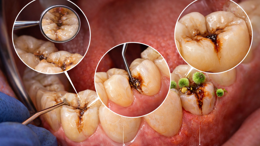
A cavity is a structural hole or defect in the tooth caused by bacterial acid erosion that breaks down the hard mineral structure of enamel and dentin. Dentists use the term dental caries interchangeably with cavities, referring to the disease process rather than just the final hole.
Decay does not start randomly across tooth surfaces. Three specific locations account for approximately 90 percent of all new cavities:
- Pits and fissures (chewing surface grooves): The deep natural grooves on molars and premolars trap food particles and bacteria that toothbrush bristles cannot reach. These grooves can be narrower than a single bristle yet extend deep into enamel, creating perfect breeding grounds for decay-causing bacteria. Children and teenagers develop 80 percent of their cavities in these grooves because newly erupted teeth have especially deep fissures.
- Interproximal areas (between teeth): The contact points where adjacent teeth touch create tight spaces where floss should pass but often does not. Plaque accumulates here undisturbed, producing acids against two tooth surfaces simultaneously. Adults develop more between-tooth cavities than children because teeth shift closer together with age, making these spaces harder to clean.
- Gingival margins (along the gumline): The border where teeth meet gums collects plaque easily, particularly when gums recede and expose softer root surfaces that lack enamel protection. Root surface cavities progress faster than enamel cavities because dentin (the layer beneath enamel) demineralizes at a higher pH (6.7) compared to enamel (5.5), meaning milder acids can cause damage.
The “Acid Attack” Cycle After Eating: What Happens in the Next 30 to 60 Minutes
Your mouth experiences a predictable acid attack lasting 30 to 60 minutes every time you eat or drink anything except plain water, during which tooth enamel temporarily softens and loses minerals. This cycle represents the fundamental mechanism of cavity formation, yet most people remain completely unaware it is happening.
The sequence unfolds in three distinct phases:
- Immediate response (0 to 5 minutes): You consume food or drink containing fermentable carbohydrates (sugars and starches). Bacteria living in plaque immediately begin metabolizing these carbohydrates, producing lactic acid, acetic acid, and other organic acids as waste products. Your mouth pH, which normally sits at a neutral 6.8 to 7.2, drops rapidly.
- Critical damage window (5 to 30 minutes): Mouth pH falls below 5.5 (the critical pH for enamel), triggering demineralization. Enamel is composed of tightly packed hydroxyapatite crystals; when surrounded by acid, calcium and phosphate ions dissolve out of the crystal structure and leach into saliva. The enamel surface becomes microscopically rougher and softer. You cannot feel this happening, and no pain signals warn you of the damage.
- Recovery phase (30 to 60 minutes): Your saliva gradually neutralizes the acids, buffering mouth pH back toward neutral. Calcium and phosphate dissolved in saliva begin depositing back into enamel (remineralization). The tooth surface hardens again. Complete pH recovery takes 30 to 60 minutes in people with healthy saliva flow, but can extend beyond 2 hours in individuals with reduced saliva production.
Demineralization vs Remineralization: When Early Decay Can Still Be Reversed
Demineralization is the loss of calcium and phosphate minerals from tooth enamel due to acid exposure, while remineralization is the redepositing of these minerals back into the enamel crystal structure through saliva, a dynamic balance that determines whether decay progresses or reverses. Understanding this balance empowers you to intervene before cavities become permanent.
The demineralization-remineralization equation operates continuously. Every acid attack shifts the balance toward mineral loss. Every recovery period shifts it toward mineral gain. Net demineralization over weeks and months produces visible decay. Net remineralization maintains or strengthens enamel.
Three factors determine which direction the balance tips:
- Acid exposure duration and frequency: Longer and more frequent acid attacks overwhelm the remineralization capacity of saliva. Your teeth can handle 3 to 4 acid challenges daily and fully recover. They cannot compensate for 8 to 12 challenges from constant snacking and sweetened beverage sipping.
- Saliva quantity and quality: Healthy saliva contains supersaturated calcium and phosphate, meaning the mineral concentration exceeds what dissolves in pure water. It actively deposits minerals into enamel when pH recovers. Reduced saliva flow (from medications, mouth breathing, or health conditions) decreases both acid buffering and mineral supply. Patients with dry mouth can experience 3 to 5 times higher cavity rates even with good hygiene.
- Fluoride presence: Fluoride converts standard hydroxyapatite crystals to fluorapatite, which resists acid dissolution and promotes faster remineralization. Low fluoride levels slow remineralization; optimal levels accelerate it. The difference becomes critical during the reversal window.
The Stages of Tooth Decay (White Spot → Enamel → Dentin → Nerve/Infection)
Tooth decay progresses through five distinct stages, from reversible enamel demineralization to irreversible pulp infection, with treatment requirements and outcomes varying dramatically depending on when dentists intervene. Understanding these stages helps you recognize when immediate action is necessary versus when remineralization attempts are appropriate.
Stage 1: White spot lesion (incipient caries):
The enamel surface remains intact but subsurface demineralization creates visible white or brown discoloration. No pain occurs because nerve endings do not extend into enamel. Diagnosis requires visual inspection under good lighting; X-rays may not yet show the lesion. Treatment focuses on remineralization through fluoride, improved hygiene, and dietary modification. Professional fluoride varnish (22,600 ppm fluoride) applied quarterly can reverse approximately 50 to 70 percent of white spot lesions over 6 to 12 months.
Stage 2: Enamel decay (caries limited to enamel):
Demineralization breaks through the enamel surface, creating a small hole that bacteria colonize. The cavity remains confined to enamel (which is 2 to 2.5 millimeters thick on chewing surfaces). Still no pain, because enamel lacks nerves. Diagnosis requires clinical examination; bitewing X-rays show radiolucent (dark) areas in enamel. Treatment involves minimally invasive composite resin filling after removing decayed tissue. Small enamel cavities typically require 30 to 45 minutes to treat. Prognosis is excellent; properly placed fillings last 7 to 15 years on average with good care.
Stage 3: Dentin decay (moderate cavity):
Decay penetrates the dentin layer beneath enamel. Dentin is softer, less mineralized, and contains microscopic tubules connecting to the tooth nerve. Decay spreads faster in dentin (3 to 6 months from enamel penetration to deep dentin) compared to enamel (12 to 24 months from white spot to enamel breakthrough). Sensitivity to cold, sweet, and sometimes chewing pressure develops because stimuli reach nerve endings through dentin tubules. X-rays clearly show decay extending into dentin. Treatment requires larger fillings, and deep cavities approaching the nerve may need protective liner placement before filling. Teeth with dentin decay face higher risk of future nerve involvement if decay was extensive.
Stage 4: Pulp involvement (deep decay, pulpitis):
Decay reaches the pulp chamber containing blood vessels and nerves. Bacteria and toxins inflame pulp tissue, causing reversible pulpitis (sharp pain that resolves quickly with stimulus removal) or irreversible pulpitis (spontaneous throbbing pain lasting hours, often worse at night or when lying down). Diagnosis involves pain history, percussion testing (tapping the tooth), cold testing, and X-rays showing decay into or near the pulp chamber. Treatment options split based on pulp vitality. Reversible pulpitis may respond to deep filling with pulp-capping medication, preserving nerve vitality.
Irreversible pulpitis requires root canal treatment (RCT) to remove infected pulp, clean canals, and seal the space. If you are unsure whether your pain has crossed this threshold, reviewing the top signs you need a root canal can help you decide whether to seek urgent care. RCT for a single-rooted tooth takes approximately 60 to 90 minutes; multi-rooted molars require 90 to 120 minutes and sometimes 2 appointments.
Stage 5: Abscess and infection:
Untreated pulpitis progresses to pulp necrosis (nerve death) and infection extending beyond the tooth into surrounding bone. Pus accumulates at the root tip, forming a periapical abscess. Symptoms include severe constant pain, swelling, fever, bad taste from pus drainage, and sometimes facial swelling requiring emergency care. If you are experiencing any of these, understanding tooth abscess symptoms and urgent treatment is critical before the infection spreads further.
X-rays show dark areas (radiolucency) around root tips indicating bone destruction. Treatment options include root canal treatment if enough tooth structure remains to support a crown, or extraction if the tooth is unsalvageable. Abscesses require antibiotic coverage (typically Amoxicillin 500 milligrams 3 times daily for 5 to 7 days) before definitive treatment. Untreated dental abscesses can spread to facial spaces, requiring hospitalization for intravenous antibiotics and surgical drainage in severe cases.
The critical lesson: treatment cost and complexity increase exponentially with each stage. A small filling costs approximately 2,000 to 4,000 Nepali Rupees (NPR) and preserves natural tooth structure, but understanding your cavity symptoms and filling options ahead of time can help you act before the situation escalates. Delaying until RCT becomes necessary costs approximately 9,800 to 16,000 NPR for the root canal plus 8,000 to 15,000 NPR for a crown (total 17,800 to 31,000 NPR). Waiting until extraction costs approximately 1,500 to 3,000 NPR for removal but 60,000 to 130,000 NPR for an implant replacement. Early detection and treatment make financial and clinical sense.
Plaque and Bacteria: Why Cavities Keep Coming Back
Plaque is a sticky biofilm that reforms quickly on teeth and shelters bacteria that feed on sugars and refined carbs. These bacteria create acids and also build a protective matrix, making plaque harder to remove if brushing and cleaning between teeth are inconsistent. Crowded teeth, braces, deep grooves, and old fillings act as “plaque traps,” which is why cavities often recur in the same high-risk areas.
What Plaque Is (Biofilm) and How Quickly It Re-Forms After Brushing
Dental plaque is a structured biofilm community of bacteria embedded in a self-produced protective matrix that adheres to tooth surfaces, reforming within 4 to 12 hours after complete removal and reaching mature, acid-producing capacity within 24 to 48 hours. Understanding plaque as a biofilm rather than simple debris explains why cavities recur even with seemingly good oral hygiene.
Plaque differs fundamentally from food debris. Food particles on teeth attract bacteria but do not directly cause decay. Plaque consists of organized bacterial colonies (over 700 different species) living in a sticky polysaccharide matrix they secrete. This biofilm architecture protects bacteria from saliva antimicrobial factors, fluoride, and even antibiotics at concentrations that kill free-floating bacteria.
How Cavity-Causing Bacteria Turn Sugar/Starch Into Acids That Weaken Enamel
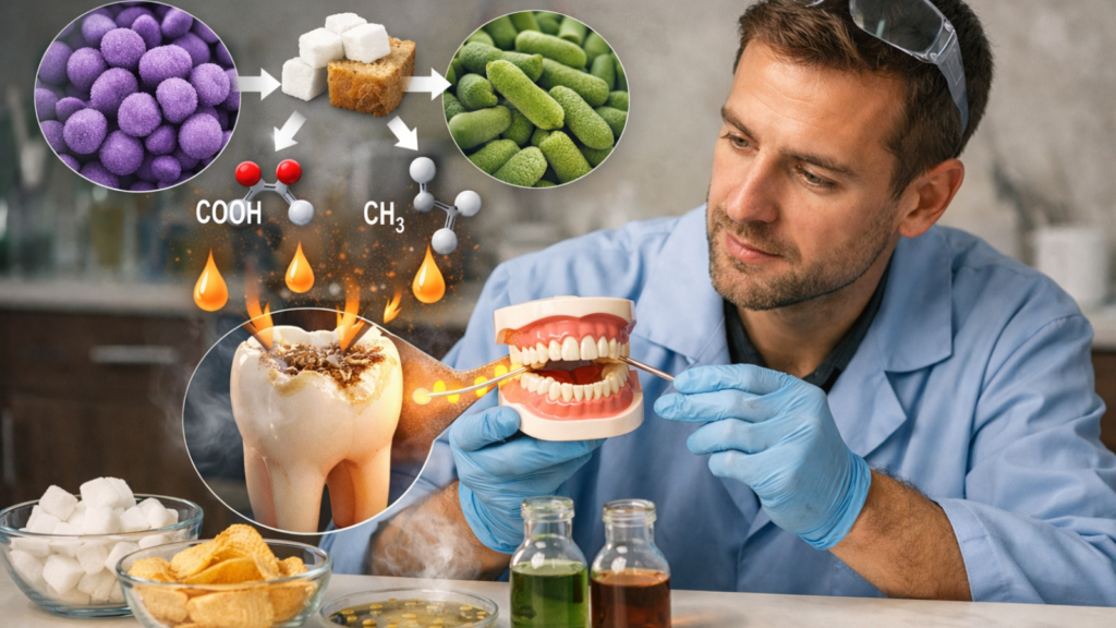
Cavity-causing bacteria, particularly Streptococcus mutans and Lactobacillus species, metabolize dietary sugars and starches through fermentation pathways that produce lactic acid, acetic acid, and other organic acids capable of dissolving tooth enamel at pH levels below 5.5. Understanding this biochemical process reveals why certain foods cause more damage than others.
The acid production mechanism operates through several steps:
- Carbohydrate uptake: Bacteria possess specific transport proteins that import simple sugars (glucose, fructose, sucrose) and break down complex starches into glucose units using enzymes called glucosyltransferases. Streptococcus mutans excels at metabolizing sucrose (table sugar), producing sticky glucan polymers that strengthen biofilm attachment while simultaneously fermenting glucose for energy.
- Fermentation pathway: Bacteria metabolize sugars through glycolysis, a multi-step chemical process that breaks down 6-carbon sugars into 3-carbon pyruvate molecules. In the low-oxygen environment within plaque biofilms, bacteria convert pyruvate into lactic acid (which lowers pH more dramatically than other acids), acetic acid, and small amounts of formic and propionic acids. This fermentation generates energy (ATP) for bacterial growth and reproduction.
- Acid accumulation: Bacteria release these acids directly into the biofilm matrix and surrounding environment. The plaque biofilm traps acids against tooth surfaces, preventing saliva from neutralizing them immediately. pH at the tooth-plaque interface drops to 4.5 to 5.0 within 5 to 10 minutes of sugar exposure, well below the critical pH of 5.5 that initiates enamel demineralization.
- Sustained acid production: Bacteria store excess sugar as intracellular polysaccharides (glycogen-like molecules). They continue fermenting these reserves and producing acids for 30 to 90 minutes after the initial sugar exposure, extending the acid attack even after you stop eating.
Different sugars and starches produce varying acid responses:
- Sucrose (table sugar): The most cavity-promoting sugar. Streptococcus mutans uses sucrose to build sticky biofilm polymers while simultaneously fermenting it for acid production. Sucrose consumption increases both biofilm thickness and acid output.
- Glucose and fructose: Metabolized rapidly, producing quick pH drops but less biofilm enhancement compared to sucrose. Fruit naturally contains fructose plus fiber and water that stimulate saliva, partially offsetting cavity risk.
- Starch (bread, rice, biscuits): Requires enzymatic breakdown into glucose before fermentation. Salivary amylase begins this process in the mouth, but bacteria complete it. Sticky starchy foods (biscuits, crackers) prolong acid production because food particles remain trapped in grooves and between teeth for hours.
- Lactose (milk sugar): Fermented slowly compared to sucrose. Pure milk poses lower cavity risk than sweetened beverages, though prolonged exposure (such as nighttime bottle feeding in children) still causes decay.
Frequency dominates quantity in determining cavity risk. Bacteria produce maximum acid within minutes of any sugar exposure, regardless of amount. Eating 10 grams of sugar once creates one acid attack. Eating the same 10 grams as 1-gram portions spread across 10 snacking occasions creates 10 separate attacks with cumulative acid exposure far exceeding the single large dose.
Kathmandu dietary patterns amplify this frequency effect. Traditional practices of frequent tea breaks with sugar and biscuits create continuous acid production throughout the day. One patient consumed sweet tea at 7 AM, 10 AM, 1 PM, 3 PM, and 6 PM, plus meals at 8 AM, 12 PM, and 7 PM (8 total acid exposures). Consolidating sweet tea to mealtimes only reduced daily acid attacks from 8 to 3, allowing adequate remineralization time and halting new cavity formation.
The specific bacterial composition matters. Not everyone harbors high levels of Streptococcus mutans. People with low S. mutans colonization can tolerate moderate sugar intake without developing cavities, while those heavily colonized develop decay rapidly even with careful diets. Mothers transmit S. mutans to infants through saliva contact (sharing spoons, pre-chewing food, mouth-to-mouth contact), establishing colonization patterns that persist lifelong.
Plaque Traps That Raise Risk: Crowded Teeth, Braces, Deep Grooves, Old Fillings
Certain dental anatomies and restorations create physical environments where plaque accumulates preferentially and resists removal, increasing cavity risk up to 3 to 5 times compared to smooth, well-aligned surfaces. Identifying your specific plaque traps allows targeted cleaning efforts and preventive interventions.
Common plaque trap categories include:
Crowded or overlapping teeth: Crowding creates tight spaces and irregular surfaces where brush bristles cannot reach. Floss sometimes cannot pass between contact points or shreds when forced through. Plaque accumulates undisturbed in these areas, producing acids that demineralize adjacent surfaces. Orthodontic treatment to align teeth improves cleanability long-term, but represents a significant investment (40,000 to 120,000 NPR for braces; 60,000 to 150,000 NPR for clear aligners in Kathmandu). Patients unable to pursue orthodontics should use interdental brushes or floss threaders specifically designed for tight contacts, plus focused fluoride application to high-risk areas.
Deep pits and fissures: Natural groove anatomy varies dramatically between individuals. Some people have shallow, easily cleaned fissures. Others possess narrow, deep grooves extending 1 to 2 millimeters into enamel, with openings smaller than a single toothbrush bristle (0.2 millimeters). Bacteria colonize deep in these fissures where mechanical cleaning cannot reach. Dental sealants (thin resin coatings applied to groove surfaces) physically block bacterial access, reducing cavity risk by approximately 70 to 80 percent over 5 years. Sealant placement costs approximately 1,000 to 2,000 NPR per tooth. The investment makes sense for children, teenagers, and adults with deep groove anatomy and a history of groove cavities.
Orthodontic appliances (braces): Fixed braces create numerous plaque retention sites around brackets, under wires, and along the gumline. Patients with braces experience 2 to 4 times higher cavity rates compared to matched controls without braces, despite similar oral hygiene efforts. The problem stems from cleaning difficulty, not lack of effort. Successful cavity prevention during orthodontic treatment requires specialized tools (orthodontic toothbrushes with V-shaped bristles, floss threaders, interdental brushes), increased cleaning time (5 to 7 minutes twice daily instead of 2 to 3 minutes), and professional fluoride applications every 3 months. White spot lesions around braces represent early decay and signal inadequate cleaning; they sometimes remain as permanent enamel scars even after braces removal.
Gingival recession exposing root surfaces: Gum recession exposes softer cementum and dentin instead of hard enamel along the gumline. These surfaces demineralize at higher pH (6.7 versus 5.5) and decay progresses approximately 2 to 3 times faster than enamel cavities. Root surface cavities often spread laterally, wrapping around the tooth circumference rather than penetrating deeply. Causes of recession include aggressive brushing, periodontal disease, thin gum tissue anatomy, and aging. For a deeper look at why this happens and whether it can be reversed, see our guide on gum recession and why gums pull back. Prevention focuses on gentle brushing technique (soft bristles, minimal pressure), gum health maintenance, and fluoride exposure to strengthen exposed surfaces.
Margins around old fillings and crowns: Dental restorations eventually develop microscopic gaps where they meet natural tooth structure due to:
- Polymerization shrinkage (composite resins contract approximately 2 to 3 percent during hardening, creating marginal gaps)
- Thermal expansion differences between restorative materials and natural teeth during hot and cold exposure
- Mechanical wear from chewing forces over years
- Incomplete margin adaptation during original placement
Bacteria colonize these gaps and produce acids that cause recurrent decay (secondary caries). Fillings typically last 7 to 15 years before requiring replacement; crowns last 10 to 20 years. Regular dental examinations catch early recurrent decay before it requires more extensive treatment.
Dry mouth conditions: Reduced saliva flow allows plaque to accumulate faster because the normal cleansing and antimicrobial effects of saliva diminish. Patients with dry mouth can develop thick, tenacious plaque within 12 to 18 hours instead of the typical 24 to 48 hours, requiring more frequent mechanical disruption (brushing after every meal instead of twice daily).
Effective management of plaque traps requires awareness and adapted technique. Many patients brush normally despite having anatomy that demands specialized approaches. Your dentist should identify your specific plaque traps during examinations and demonstrate appropriate cleaning methods. You can then focus effort where it matters most rather than using generic brushing recommendations that may miss your highest-risk areas.
Common Cavity “Types” by Location: Pit-and-Fissure, Between-Teeth, Root, Recurrent
Dentists classify cavities by location into four major categories (pit-and-fissure, interproximal, root surface, and recurrent decay), each with distinct risk factors, detection methods, and prevention strategies. Understanding which cavity types affect you most helps target prevention efforts effectively.
Pit-and-fissure cavities (occlusal caries):
These develop in the natural grooves and pits on chewing surfaces of molars and premolars. Pit-and-fissure cavities account for approximately 80 to 90 percent of cavities in children and teenagers but decrease in proportion (to approximately 40 to 50 percent of new cavities) in adults as other locations become more vulnerable.
Risk factors include deep groove anatomy, inadequate sealant protection, and difficulty cleaning grooves mechanically. Diagnosis requires visual inspection (dark or discolored grooves) and sticky explorer sensation (the dental tool catches in softened enamel). Early groove cavities sometimes evade X-ray detection because the overlying enamel masks underlying dentin decay.
Prevention centers on dental sealants for high-risk individuals. Sealants work best when applied shortly after teeth erupt, before decay starts (age 6 to 7 for first molars; age 11 to 13 for second molars). Adults with deep grooves and recurrent groove decay also benefit from sealants. Sealed teeth develop approximately 80 percent fewer cavities over 5 years compared to unsealed teeth.
Interproximal cavities (between-teeth decay):
These occur on the contact surfaces where adjacent teeth touch, accessible only by flossing or interdental cleaning. Interproximal cavities become more common with age, representing approximately 10 to 20 percent of childhood cavities but 40 to 50 percent of adult cavities.
Risk factors include tight contacts, crowding, failure to floss regularly, and between-tooth plaque accumulation. Diagnosis relies heavily on bitewing X-rays (which show cavities between teeth not visible during clinical examination) taken every 12 to 24 months based on cavity risk. Early interproximal cavities appear as small dark triangular areas on X-rays just beneath the contact point.
Prevention requires daily flossing or interdental brush use. Many patients brush regularly but never floss, leaving between-tooth surfaces completely unprotected. Proper flossing technique involves curving the floss in a C-shape around each tooth surface and sliding below the gumline, then repeating for the adjacent tooth surface. The process requires approximately 2 to 3 minutes to clean all interproximal surfaces thoroughly.
Root surface cavities (root caries):
These develop on exposed root surfaces near or below the gumline where enamel protection is absent. Root cavities typically affect adults over 40 years old, particularly those with gum recession, and account for approximately 10 to 15 percent of adult cavities.
Risk factors include gum recession (from periodontal disease or aggressive brushing), dry mouth, high sugar exposure, and aging (root caries incidence doubles every decade after age 50). Root surfaces covered only by thin cementum (a softer mineral layer) and underlying dentin demineralize at pH 6.7 instead of the enamel threshold of 5.5, making them vulnerable to weaker acids.
Diagnosis involves examining areas of gum recession for soft, discolored patches. Root cavities often spread laterally along the root circumference rather than penetrating deeply, creating shallow, wide lesions that can encircle the tooth. Treatment can be challenging because root surface access is difficult and restorations in this area face high failure rates (approximately 20 to 30 percent over 5 years).
Prevention emphasizes fluoride exposure (professional varnish application every 3 months; daily fluoride mouthrinse), gentle brushing to prevent further recession, and aggressive sugar exposure reduction. Patients with multiple root cavities may benefit from prescription-strength fluoride toothpaste (5,000 ppm fluoride) used daily.
Recurrent decay (secondary caries):
This represents new cavities forming at the margins of existing fillings or crowns. Recurrent decay accounts for approximately 50 to 60 percent of restorative dentistry procedures (most dentist time is spent replacing old restorations, not treating primary cavities).
Risk factors include marginal gaps in old restorations, inadequate oral hygiene around margins, high sugar exposure, and dry mouth. Large, complex restorations face higher recurrent decay rates than small, simple fillings. Diagnosis involves visual inspection of restoration margins, X-rays showing radiolucency (dark areas) adjacent to restorations, and explorer sensation of softness.
Prevention requires recognizing that fillings and crowns are not permanent. They require replacement every 7 to 20 years depending on size, material, location, and your cavity risk. Maintaining excellent oral hygiene and professional monitoring allows dentists to detect early recurrent decay and replace restorations before extensive tooth structure is lost. Delaying replacement of failing restorations often results in larger decay requiring more aggressive treatment (such as crown instead of filling; root canal instead of crown).
Understanding your personal cavity pattern helps optimize prevention. Someone who develops mostly groove cavities benefits most from sealants. Another person with predominantly between-teeth decay needs better flossing technique and possibly fluoride focus on interproximal surfaces. Patients with recurrent decay around old fillings should question whether underlying risk factors (diet, dry mouth, systemic health) have changed and address those issues instead of repeatedly replacing restorations without modifying contributing factors.
Diet Patterns That Accelerate Cavities (and What to Change)

For cavities, how often you expose teeth to sugar matters as much as (or more than) how much you eat, because each snack triggers a new acid attack. Sticky snacks and sweetened drinks, especially sipped slowly, keep the mouth acidic longer and give enamel less time to recover. Simple changes like limiting sugary items to mealtimes, choosing water between meals, and rinsing after sweets can reduce the total acid time each day.
Sugar Frequency vs Sugar Amount: Why Snacking/Sipping All Day Is Worse
Cavity risk correlates more strongly with the frequency of sugar exposures throughout the day than with the total quantity of sugar consumed, because each exposure triggers a 30 to 60 minute acid attack regardless of the amount ingested. This counterintuitive principle explains why some people who consume significant sweets develop few cavities while others with seemingly modest sugar intake experience rampant decay.
The frequency-versus-amount concept rests on the acid attack duration principle explained earlier. Your mouth pH drops below the critical threshold (5.5) within minutes of any sugar exposure and remains acidic for 30 to 60 minutes until saliva buffers it back to neutral. Total acid exposure time depends on attack frequency, not sugar quantity per attack.
The solution focuses on timing, not elimination:
- Consolidate sweet intake to mealtimes: Desserts eaten immediately after meals do not create separate acid attacks. The meal already triggered acid production; adding sweets extends the attack slightly but does not multiply frequency. Eating the same dessert 2 hours after the meal doubles attack frequency.
- Limit between-meal snacking: Restrict eating to defined meal times (3 to 4 daily maximum). It gets easier to maintain healthy pH when acid attacks are predictable and spaced.
- Switch to unsweetened beverages between meals: Plain water, unsweetened tea, black coffee (without sugar) do not trigger acid production. Reserve sweetened versions for mealtimes only.
- End meals with cheese or nuts: Cheese (particularly aged varieties) and nuts stimulate saliva production and help buffer acids. Some evidence suggests they may promote remineralization. Ending a meal with a small piece of cheese instead of sweets slightly reduces net acid exposure.
This approach allows enjoyment of sweets while dramatically reducing cavity risk. You can consume a moderate amount of sugar with minimal tooth damage by controlling timing. Conversely, even small sugar amounts distributed throughout the day cause significant harm.
Many patients resist this advice initially, viewing frequent tea or snacking as necessary for energy or social connection. However, pilot trials in Kathmandu clinics show that consolidating sweet tea to 2 to 3 times daily (instead of 5 to 6) reduces new cavity formation by approximately 40 to 60 percent over 12 to 18 months while maintaining patient satisfaction. The adjustment period lasts approximately 1 to 2 weeks; most patients adapt readily and report no loss of enjoyment.
Hidden Sugars and Sticky Carbs: Biscuits, Sweetened Tea/Coffee, Juices, Packaged Snacks
Many Kathmandu dietary staples contain high levels of hidden sugars or sticky refined carbohydrates that prolong acid exposure, contributing to cavity formation even in patients who believe they consume minimal sweets. Identifying these sources and understanding their impact enables practical dietary modifications that preserve cultural food preferences while reducing decay risk.
Common hidden sugar and sticky carbohydrate sources include:
- Biscuits and packaged cookies: Popular brands widely available in Kathmandu contain 20 to 35 percent sugar by weight plus refined flour that breaks down to glucose. The sticky texture causes particles to adhere to grooves and between teeth for 30 to 90 minutes after consumption, providing prolonged sugar exposure to bacteria. A single serving (3 to 4 biscuits) delivers approximately 10 to 15 grams sugar. Someone consuming biscuits during morning and afternoon tea breaks ingests 20 to 30 grams from this source alone, not counting sugar added to tea.
- Sweetened tea and coffee: Traditional sweet tea preparation adds 1 to 2 teaspoons (4 to 8 grams) sugar per cup. Individuals consuming 4 to 6 cups daily ingest 16 to 48 grams of sugar just from beverages, delivered in frequent small doses that maximize acid attack frequency. Instant coffee mixes and packaged tea drinks often contain even more sugar (6 to 10 grams per serving) than home-prepared versions.
- Fruit juices and sweetened drinks: Packaged juices contain 10 to 15 grams sugar per 200 milliliter serving, comparable to sodas. Even “100 percent fruit juice” lacks the fiber present in whole fruit and concentrates natural sugars. Drinking juice causes pH to drop just as dramatically as drinking soda. Mango lassi, popular in local tea shops, typically contains added sugar beyond the natural milk sugar, delivering 15 to 25 grams per serving.
- Flavored milk and yogurt drinks: Marketed as healthy options, these products often contain 12 to 20 grams added sugar per 200 milliliter serving. Patients switching from soda to flavored milk receive minimal cavity prevention benefit despite perceiving a healthier choice.
- Traditional sweet snacks: Sel roti, jalebi, laddu, and similar festival or everyday treats combine high sugar content (30 to 50 percent by weight) with sticky textures that prolong tooth contact time. Sel roti’s porous structure traps between teeth particularly effectively.
- Dried fruits: Marketed as natural and healthy, dried fruits (raisins, dates, dried mango) concentrate natural sugars and develop extremely sticky textures that adhere to teeth for hours. Decay potential rivals traditional candy. Whole fresh fruit contains identical sugar but includes water and fiber that stimulate saliva and reduce adhesion, making it significantly less cavity-promoting.
Reading ingredient labels reveals hidden sugars in unexpected products. Sugar appears under numerous names: sucrose, glucose, fructose, dextrose, maltose, corn syrup, cane juice, honey, molasses, and many others. Products listing sugar (in any form) among the first 3 to 4 ingredients contain substantial amounts.
The sticky carbohydrate problem deserves particular attention. Starchy foods that adhere to teeth provide prolonged bacterial fuel even without high direct sugar content. Examples include:
- White bread and crackers
- Potato chips and fried snacks
- Many breakfast cereals
- Sticky rice varieties
These foods break down to glucose through salivary amylase activity and bacterial enzymes, then ferment into acids. The prolonged contact time (from sticky adherence) extends acid production for 60 to 120 minutes or more.
Practical modifications that maintain cultural food patterns while reducing cavity risk:
For tea culture: Reduce sugar concentration gradually (from 2 teaspoons to 1.5, then 1, then 0.5 over 4 to 6 weeks). Reduce frequency (from 6 cups to 4, then 3). Reserve sweet tea for morning and mid-afternoon only; drink unsweetened tea or water at other times. Many patients find they enjoy tea flavor more clearly with less sugar after the adjustment period.
For snacking: Replace biscuits with less sticky options (nuts, cheese, fresh fruit, roasted chickpeas). These alternatives satisfy hunger and social eating without creating prolonged acid exposure. Rinse mouth with water immediately after consuming sticky snacks when brushing is impractical.
For beverages: Switch to water as the primary between-meal drink. Limit juice to small portions (100 to 150 milliliters) consumed with meals, not sipped throughout the day. Dilute juice with water (50:50 ratio) when provided to children.
For traditional sweets: Enjoy them during festivals and special occasions (maintaining cultural practice) but avoid incorporating them into daily routine. When consumed, eat them with or immediately after meals rather than as standalone snacks.
These modifications require minimal lifestyle disruption but produce measurable cavity reduction. Clinic data from Kathmandu patients show that reducing sweet tea frequency from 5 to 6 cups daily to 2 to 3 cups, plus replacing afternoon biscuits with nuts or cheese, reduces new cavity formation by approximately 50 percent over 12 months. Patients report the changes feel manageable after initial adjustment.
Cavities vs Enamel Erosion: How Acidic Drinks Damage Teeth Differently (and Together)
Cavities result from bacterial acid production through sugar fermentation, while enamel erosion occurs through direct chemical dissolution by acidic beverages and foods, two distinct damage mechanisms that often act synergistically to accelerate tooth destruction. This is especially relevant for people who experience acid reflux, as acid reflux and enamel erosion can compound cavity risk significantly. Understanding this difference helps explain why some dietary patterns cause widespread enamel loss beyond typical cavity patterns.
Key distinctions between cavities and erosion:
Mechanism:
- Cavities: bacteria metabolize sugar → produce acids → demineralize specific localized areas creating holes
- Erosion: acidic foods/drinks directly contact enamel → dissolve minerals across broad surfaces → thin enamel uniformly
Location:
- Cavities: pits/fissures, between teeth, gumline, around restorations (wherever plaque accumulates)
- Erosion: front surfaces of front teeth, chewing surfaces of all teeth, anywhere repeated acid contact occurs
Pattern:
- Cavities: well-defined holes with bacterial infection present
- Erosion: generalized wearing, thinning, cupping of chewing surfaces; smooth, shiny appearance; transparency of edges
Prevention:
- Cavities: reduce sugar frequency, improve plaque removal, increase fluoride exposure
- Erosion: limit acidic beverage exposure, use straws to bypass teeth, avoid brushing immediately after acid exposure
Common erosive beverages and foods include:
- Soft drinks and sodas: pH 2.5 to 3.5 (highly erosive). Cola drinks contain phosphoric acid; citrus sodas contain citric acid. Both dissolve enamel on contact. One patient who consumed 1 to 2 liters of cola daily for 5 years presented with severe erosion (enamel completely worn through to dentin on all front teeth), requiring full-mouth crowns costing approximately 400,000 to 600,000 NPR.
- Sports and energy drinks: pH 2.9 to 3.2. Marketed as healthy hydration but highly acidic. Frequent consumption during exercise (when mouth is dry and saliva buffering is minimal) causes severe damage.
- Fruit juices: Orange juice pH 3.3 to 4.2; apple juice pH 3.4 to 4.0. Natural fruit acids (citric, malic, tartaric) erode enamel despite absence of added sugars. Prolonged sippy-cup use in toddlers (allowing juice contact for hours) causes devastating erosion.
- Kombucha and fermented drinks: pH 2.5 to 3.5 due to acetic acid from fermentation. Growing popularity in urban Kathmandu increases erosion risk.
- Vinegar-based foods: Pickles, salad dressings, certain chutneys. Repeated consumption contributes to erosion, particularly when held in mouth.
- Wine (particularly white wine): pH 3.0 to 3.5. Wine tasting (swishing and holding) produces severe erosion in front teeth. Red wine pH is similar but tannins provide slight protective effect.
- Citrus fruits: Lemons pH 2.2 to 2.4; oranges pH 3.7 to 4.3. Eating whole fruit causes less erosion than juice because chewing time is shorter. Sucking on lemons (a practice some adopt for perceived health benefits) causes extreme focal erosion.
The synergistic cavity-erosion problem arises when drinks contain both sugar (promoting bacterial acid production) and intrinsic acidity (causing direct erosion). Soft drinks represent the worst offenders, delivering double damage. Each sip triggers immediate erosive demineralization from drink pH 2.5 to 3.5, then prolonged bacterial acid production as sugar ferments.
Practical Tooth-Friendly Habits: Timing Sweets, Rinsing With Water, Smarter Snack Swaps
Simple behavioral modifications in eating timing, beverage choices, and post-consumption practices can reduce cavity risk by 40 to 70 percent without requiring complete dietary overhaul or elimination of enjoyed foods. These evidence-based strategies work by minimizing acid exposure duration and enhancing natural protective mechanisms.
Timing strategies that reduce acid exposure:
- Consume sweets and acidic items only during main meals: Desserts, sweetened beverages, and acidic drinks create minimal additional acid exposure when consumed immediately after meals (which already triggered acid production). The same items consumed 2 to 3 hours after meals create entirely separate acid attacks. Consolidating sweets to mealtimes can reduce daily acid attacks from 7 to 8 down to 3 to 4, cutting total acid exposure time by approximately 50 percent.
- End meals with protective foods: Cheese (particularly aged varieties like cheddar, Swiss, Gouda) stimulates saliva production and contains calcium and phosphate that buffer acids and support remineralization. Finishing a meal with a small cheese portion (20 to 30 grams) reduces the severity and duration of post-meal acid attacks. Nuts (almonds, cashews, peanuts) provide similar benefits through stimulating saliva flow and delivering minimal fermentable carbohydrate. These foods also satisfy the desire for a distinct meal ending without triggering additional acid exposure.
- Avoid eating or drinking anything except water for 1 to 2 hours before bed: Saliva production decreases dramatically during sleep (up to 90 percent reduction compared to waking flow). Acids linger much longer without buffering, and teeth remain in demineralizing pH for extended periods. A bedtime snack creates an acid attack that persists for 2 to 4 hours instead of the typical 30 to 60 minutes, causing significant damage. Water remains the only safe pre-bed beverage.
Immediate post-eating practices:
- Rinse mouth with plain water for 30 seconds: This simple practice (done immediately after eating sweets, acidic foods, or sticky carbohydrates) provides multiple benefits. Water dilutes residual sugars and acids, physically washes away loose food particles, and helps restore neutral pH faster. Swishing water vigorously for 30 seconds removes approximately 30 to 40 percent of fermentable material compared to doing nothing. The practice costs nothing, requires no special products, and works anywhere.
- Chew sugar-free gum for 5 to 10 minutes (xylitol preferred): Chewing stimulates saliva flow 5 to 10 times above resting levels, dramatically accelerating acid buffering and remineralization. Sugar-free gum sweetened with xylitol provides additional benefits because xylitol cannot be fermented by cavity-causing bacteria, and regular xylitol exposure (5 to 10 grams daily) reduces Streptococcus mutans levels in plaque over several months. Gum chewing immediately after meals or snacks reduces cavity risk by approximately 20 to 30 percent in controlled studies. Choose products containing xylitol as the first ingredient (indicating primary sweetener status).
- Delay brushing 30 to 60 minutes after consuming acidic items: Brushing immediately after acidic beverage or food consumption (citrus, soda, wine, vinegar-based items) mechanically abrades softened enamel, accelerating erosion. Wait 30 to 60 minutes to allow saliva to reharden enamel, then brush. For non-acidic meals, brushing immediately poses no erosion risk.
Smart snack substitutions that reduce cavity risk:
Replace high-risk snacks with lower-risk alternatives that satisfy similar cravings without prolonged acid exposure:
Instead of biscuits or cookies → choose nuts (almonds, cashews, peanuts), cheese cubes, or roasted chickpeas: These options deliver satisfaction and satiety with minimal fermentable carbohydrate. Nuts and cheese actually promote remineralization rather than demineralization.
Instead of dried fruit → choose fresh whole fruit: Fresh fruit contains identical natural sugars but includes water that dilutes sugars, fiber that stimulates saliva, and shorter eating duration (you finish an apple in 5 to 10 minutes versus hours of nibbling sticky raisins). The cavity risk difference is substantial despite similar sugar content.
Instead of sweetened yogurt drinks → choose plain yogurt with fresh fruit added at home: Commercial fruit yogurts and drinks contain 12 to 20 grams added sugar. Plain yogurt with fresh berries delivers similar taste with approximately 50 to 60 percent less total sugar and zero added sugars. The bacterial cultures (probiotics) in yogurt may provide additional oral health benefits through competing with cavity-causing bacteria.
Instead of juice → choose whole fruit with water: This substitution eliminates the concentrated sugar exposure of juice while maintaining fruit intake. An orange contains approximately 12 grams sugar consumed in 5 to 10 minutes; 200 milliliters of orange juice contains the sugar of 2 to 3 oranges consumed in seconds and often sipped over 30 to 60 minutes.
Instead of sweetened tea/coffee → gradually reduce sugar then eliminate: Many patients successfully reduce tea sugar from 2 teaspoons to zero over 4 to 8 weeks through gradual 0.25 teaspoon decrements every 7 to 10 days. The taste adjustment occurs gradually, and most report preferring less-sweet tea after adaptation.
Instead of soft drinks → choose sparkling water, herbal tea, or milk: Plain sparkling water (pH 5.0 to 5.5) is mildly acidic but lacks sugar and poses minimal cavity risk. It satisfies the desire for carbonation and flavor without promoting decay. Unsweetened herbal teas provide flavor variety. Milk delivers calcium and phosphate that support remineralization.
Hydration optimization:
Maintain adequate water intake (2 to 3 liters daily): Proper hydration supports optimal saliva production. Dehydration reduces saliva flow, allowing plaque to accumulate faster and acids to persist longer. Water intake should increase in hot weather, during illness, and with physical activity. Patients taking multiple medications (which often cause dry mouth as a side effect) require particularly vigilant hydration.
Sip water throughout the day: Keeping water readily available and sipping small amounts every 15 to 30 minutes maintains saliva production and mechanically cleanses teeth. This habit becomes especially important for people with dry mouth tendencies.
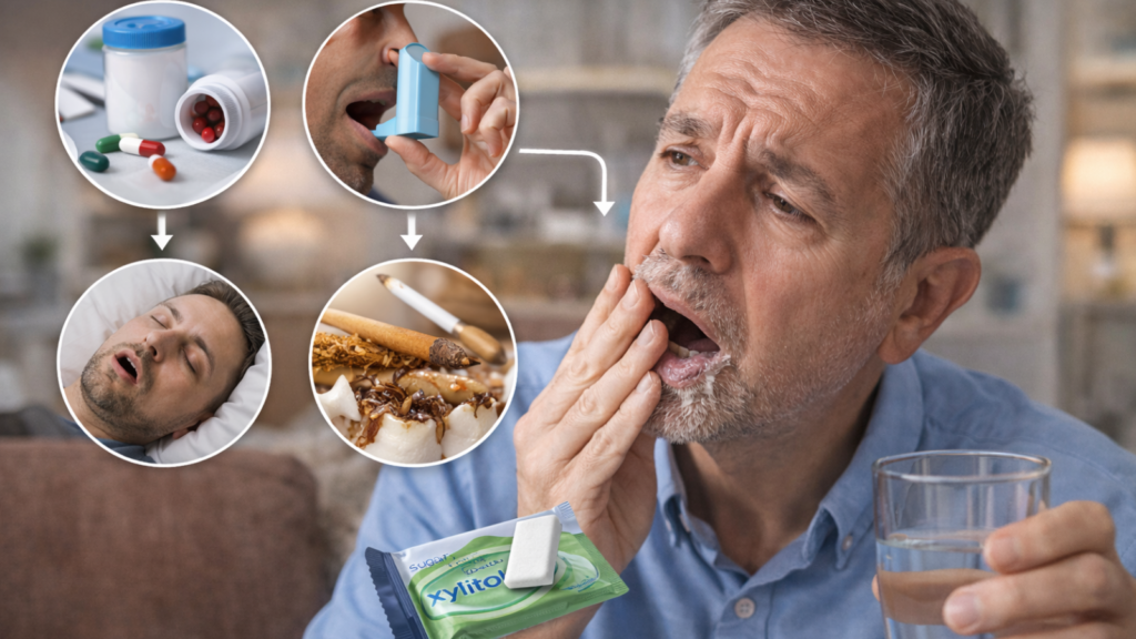
Dry Mouth (Low Saliva): A Major Risk Factor People Miss
Saliva protects teeth by washing away food, buffering acids, and helping remineralize early damage, so low saliva makes cavities form faster and spread more widely. Dry mouth can come from dehydration, mouth breathing, tobacco, and many common medications, and it often shows up as frequent thirst, sticky saliva, bad breath, or new gumline/root cavities. Improving hydration, using sugar-free gum (often with xylitol), and getting dentist-guided fluoride support can make a big difference when dryness is persistent.
Why Saliva Protects Teeth: Buffering Acids, Washing Plaque, Rebuilding Enamel
Saliva serves as the mouth’s primary natural defense system against tooth decay through three critical mechanisms: neutralizing bacterial acids, mechanically cleansing food debris and plaque, and providing minerals for enamel remineralization. Reduced saliva flow eliminates these protective effects and increases cavity risk by 3 to 5 times compared to normal saliva production, yet many patients and healthcare providers overlook dry mouth as a significant factor.
The three primary protective mechanisms operate continuously in healthy individuals:
Acid buffering and pH regulation:
Saliva contains bicarbonate, phosphate, and protein buffering systems that neutralize acids produced by bacteria or consumed in foods and beverages. Resting saliva maintains mouth pH at approximately 6.8 to 7.2 (near neutral). Bacterial acid production or acidic beverage consumption drops pH toward 4.5 to 5.5 within minutes. Saliva buffering gradually restores neutral pH over 30 to 60 minutes through chemical neutralization reactions.
Healthy saliva flow (1 to 1.5 milliliters per minute when stimulated; 0.3 to 0.5 milliliters per minute at rest) delivers sufficient buffering capacity to neutralize typical acid challenges within 30 to 60 minutes. Reduced flow (below 0.1 milliliters per minute at rest; below 0.7 milliliters per minute stimulated) cannot buffer adequately, allowing pH to remain acidic for 90 to 180 minutes or longer. This extended acid exposure dramatically increases demineralization and cavity formation.
The buffering effect becomes particularly critical during sleep. Saliva production decreases approximately 90 percent during sleep compared to waking levels. People with already-reduced saliva face near-zero flow during sleep, creating 6 to 8 hours of minimal acid buffering. Bedtime snacks or sweetened beverages become especially destructive in dry-mouth patients.
Mechanical cleansing action:
Saliva flow continuously washes food particles, bacterial debris, and soluble sugars from tooth surfaces and oral soft tissues. Swallowing occurs approximately 2,000 times daily in healthy adults, each swallow clearing accumulated material from the mouth. This mechanical cleansing reduces substrate availability for bacterial fermentation and physically disrupts biofilm formation.
Dry mouth patients report food sticking to teeth, difficulty swallowing, and thick, ropy saliva (when reduced flow concentrates mucus proteins). The cleansing deficit allows plaque to accumulate much faster. Studies show dry mouth patients develop mature, acid-producing biofilms in 12 to 18 hours instead of 24 to 48 hours, effectively doubling the required brushing frequency to maintain cavity control.
The cleansing function extends beyond teeth to include tongue, cheeks, and throat. Reduced flow allows bacterial overgrowth on the tongue (visible as thick white or yellow coating), contributes to bad breath (halitosis), and increases throat infections. Many dry mouth patients develop chronic oral candidiasis (fungal infection presenting as white patches or burning sensation).
Mineral supply for remineralization:
Healthy saliva is supersaturated with calcium and phosphate ions, meaning it contains higher mineral concentrations than would normally dissolve in water. Saliva delivers these ions directly to tooth surfaces, where they deposit into demineralized enamel during pH recovery periods following acid attacks. This remineralization process repairs early cavity damage before it progresses to irreversible holes.
Specific salivary proteins (statherin, proline-rich proteins, histatins) regulate mineral deposition and prevent enamel from dissolving even when pH approaches the critical threshold. These proteins maintain a protective film on tooth surfaces.
Reduced saliva flow decreases mineral delivery and removes protein protection. Demineralized areas cannot repair effectively. White spot lesions progress to cavitation instead of reversing. Even with good oral hygiene, dry mouth patients experience cavity formation because the natural remineralization mechanism fails.
Causes of Dry Mouth: Medications, Dehydration, Mouth Breathing, Tobacco, Health Conditions
Dry mouth (xerostomia) results from numerous medical, behavioral, and environmental factors, with medication side effects representing the most common cause in adult populations, affecting approximately 400 to 600 commonly prescribed drugs including antihypertensives, antidepressants, antihistamines, and pain medications. Identifying and when possible modifying these causes becomes essential for cavity prevention in affected individuals.
1. Medication-induced dry mouth:
Over 500 medications list dry mouth as a documented side effect. The following categories produce xerostomia most frequently:
Antihypertensives (blood pressure medications): Diuretics (hydrochlorothiazide, furosemide) reduce overall body fluid volume, decreasing saliva production. ACE inhibitors, calcium channel blockers, and beta-blockers affect salivary gland function through various mechanisms. Approximately 10 to 30 percent of patients on these medications experience noticeable dry mouth.
Antidepressants and anti-anxiety medications: Tricyclic antidepressants (amitriptyline, nortriptyline) cause severe dry mouth in 60 to 80 percent of users. SSRIs (fluoxetine, sertraline, escitalopram) produce dry mouth in 15 to 30 percent of patients. Benzodiazepines (diazepam, alprazolam) affect approximately 10 to 20 percent. The anticholinergic effects of these medications directly block saliva secretion.
Antihistamines (allergy medications): Both prescription and over-the-counter antihistamines (cetirizine, loratadine, diphenhydramine) reduce saliva through anticholinergic action. Approximately 20 to 40 percent of users notice dry mouth.
Pain medications: Opioid analgesics (tramadol, codeine, morphine) produce dry mouth in 30 to 50 percent of users. Non-steroidal anti-inflammatory drugs (ibuprofen, naproxen) cause dry mouth less frequently (approximately 5 to 10 percent).
Others: Antipsychotics, muscle relaxants, anti-nausea medications, bladder control drugs, and many others contribute. Patients taking multiple medications from different categories experience cumulative dry mouth effects.
Management requires collaboration with prescribing physicians. Options include:
- Switching to alternative medications within the same class that cause less dry mouth (for instance, switching from a tricyclic antidepressant to an SSRI with lower anticholinergic activity)
- Reducing dosages when medically appropriate
- Timing medication doses to minimize dry mouth impact (for instance, taking medications causing severe dry mouth in the morning rather than bedtime when saliva flow is already minimal)
- Balancing medication benefits against dry mouth risks (stopping essential cardiac medications is not appropriate; managing dry mouth through other means becomes necessary)
Never discontinue prescribed medications without physician consultation. Managing dry mouth through other interventions while continuing necessary medications often represents the appropriate approach.
2. Dehydration:
Inadequate fluid intake reduces total body water content, decreasing saliva production. Chronic mild dehydration affects many individuals without obvious symptoms. Signs include dark concentrated urine, infrequent urination (less than 4 to 6 times daily), fatigue, and mild headaches.
Dehydration risk increases with:
- Hot climate (Kathmandu summer temperatures often exceed 30 to 35 degrees Celsius)
- Physical activity without adequate fluid replacement
- Vomiting, diarrhea, or fever
- Excessive caffeine or alcohol consumption (both promote fluid loss)
- Deliberate fluid restriction (some patients drink minimally to avoid frequent urination)
Adequate hydration requires approximately 2 to 3 liters total fluid daily for adults (more during hot weather or exercise). Plain water represents the ideal fluid. Sweetened beverages contribute to hydration but introduce cavity-promoting sugars, creating counterproductive effects.
Patients with medication-induced dry mouth require extra attention to hydration because adequate fluid intake partially compensates for reduced saliva production. Inadequate hydration plus medications produces severe xerostomia and rampant decay.
3. Mouth breathing:
Breathing primarily through the mouth (instead of the nose) causes evaporative drying of saliva and reduces its protective effects even when production is normal. Chronic mouth breathing develops from:
- Nasal congestion (allergies, chronic rhinitis, sinusitis, deviated septum)
- Enlarged adenoids or tonsils (particularly in children)
- Habitual behavior (some people mouth breathe without physiological obstruction)
- Sleep-disordered breathing (snoring, sleep apnea)
Mouth breathing particularly impacts nighttime saliva protection because it compounds the normal sleep-related flow reduction. Patients often report waking with extremely dry mouth, bad breath, and thick saliva.
Diagnosis involves observing breathing patterns at rest and during sleep (partners often report open-mouth sleeping), examining nasal passages for obstruction, and evaluating for sleep apnea symptoms (snoring, witnessed breathing pauses, morning headaches, daytime sleepiness).
Treatment addresses underlying causes (allergy management, surgical correction of anatomical obstructions, CPAP therapy for sleep apnea) and behavioral modification (conscious nasal breathing practice, mouth taping during sleep in select cases under medical supervision).
4. Tobacco and alcohol use:
Smoking and smokeless tobacco reduce saliva production through multiple mechanisms (heat damage to salivary glands, chemical effects of nicotine and other compounds, chronic inflammation). Approximately 50 to 70 percent of regular tobacco users experience dry mouth. Tobacco cessation typically improves saliva flow within 2 to 6 weeks.
Alcohol produces acute temporary dry mouth (through dehydration and direct tissue effects) and chronic dry mouth with long-term heavy consumption (through glandular damage). Even moderate social drinking creates temporary xerostomia lasting several hours.
5. Health conditions affecting salivary glands:
Several systemic diseases directly impair saliva production:
Sjögren syndrome: An autoimmune disease where the immune system attacks moisture-producing glands, particularly salivary and tear glands. Patients experience severe dry mouth and dry eyes. Sjögren syndrome affects approximately 0.5 to 1 percent of the population, predominantly middle-aged women. Diagnosis requires specific blood tests (anti-SSA, anti-SSB antibodies) and sometimes salivary gland biopsy. No cure exists; treatment focuses on symptom management and preventing complications.
Diabetes: Both type 1 and type 2 diabetes increase dry mouth risk through dehydration (from high blood glucose causing excessive urination) and neuropathy affecting salivary gland nerves. Approximately 40 to 50 percent of diabetics report dry mouth. Blood glucose control improves saliva flow; poorly controlled diabetes worsens xerostomia.
Thyroid disorders: Hypothyroidism (underactive thyroid) commonly causes dry mouth. Treatment with thyroid hormone replacement typically resolves the symptom.
HIV/AIDS: Both the infection and medications used to treat it cause reduced saliva flow.
Radiation therapy: Head and neck cancer treatment involving radiation to salivary glands produces severe, often permanent dry mouth. Radiation damages gland tissue irreversibly. Patients may produce less than 10 percent of normal saliva flow after treatment.
Chemotherapy: Many chemotherapy drugs temporarily reduce saliva production, typically recovering several weeks after treatment completion.
Aging: Salivary gland function declines modestly with aging (approximately 25 to 30 percent reduction from age 30 to 80), though severe dry mouth in elderly patients usually results from medications and health conditions rather than aging alone.
Understanding the cause guides appropriate intervention. Medication-induced dry mouth requires physician consultation about alternatives. Dehydration demands increased fluid intake. Mouth breathing needs ENT evaluation. Sjögren syndrome requires rheumatology care plus aggressive dental prevention. Identifying and addressing underlying causes when possible produces better outcomes than only treating dry mouth symptoms.
Signs of Xerostomia: Thirst, Burning Mouth, Bad Breath, Frequent New Cavities
Dry mouth manifests through multiple symptoms beyond simple thirst, including burning sensations, altered taste, difficulty swallowing, oral infections, and dramatically accelerated cavity formation, yet many patients fail to connect these seemingly unrelated problems to reduced saliva flow. Recognizing the full symptom pattern enables earlier diagnosis and intervention.
Common xerostomia symptoms include:
1. Persistent thirst and desire to sip fluids:
Patients report needing water constantly, keeping water bottles readily available, and waking multiple times nightly to drink. The chronic thirst develops because reduced saliva creates uncomfortable mouth dryness that prompts fluid seeking. Many patients sip sweetened beverages throughout the day to relieve dryness, inadvertently worsening cavity risk through continuous sugar exposure.
2. Burning mouth sensation:
Approximately 30 to 50 percent of dry mouth patients experience burning, tingling, or scalding sensations affecting the tongue, palate, lips, or entire mouth. The burning typically worsens throughout the day and may interfere with eating spicy or acidic foods. The mechanism involves reduced saliva lubrication plus altered oral microbiome that produces irritating metabolites. Burning mouth syndrome sometimes occurs without obvious dry mouth, complicating diagnosis.
3. Altered taste perception:
Dry mouth patients report metallic taste, constant bad taste, or difficulty tasting foods normally. Saliva dissolves taste molecules and delivers them to taste receptors; reduced flow impairs this process. Additionally, oral bacterial overgrowth produces compounds that create unpleasant tastes.
4. Difficulty swallowing dry foods:
Patients need frequent water sips to swallow crackers, bread, rice, and other dry foods. Some avoid these foods entirely due to swallowing difficulty. The problem stems from inadequate saliva to form a moist food bolus that slides easily through the throat.
5. Speech difficulty:
Saliva lubricates mouth tissues and enables smooth tongue and lip movements required for clear speech. Dry mouth causes lips to stick to teeth, tongue to stick to the palate, and clicking sounds during speech. Public speakers, teachers, and singers notice these issues particularly.
6. Oral infections:
Candidiasis (oral thrush): Fungal infection presenting as white patches on tongue, inner cheeks, or palate (that wipe off leaving red raw areas), or as redness and burning without visible patches (atrophic candidiasis). Approximately 30 to 40 percent of dry mouth patients develop chronic or recurrent candidiasis because reduced saliva removes antifungal protection.
Angular cheilitis: Painful cracks or splits at mouth corners, often with candidal or bacterial infection. Common in dry mouth patients and denture wearers.
Increased gum disease: Reduced saliva cleansing allows bacterial plaque to accumulate faster and more extensively, accelerating gingivitis and periodontitis.
7. Bad breath (halitosis):
Chronic dry mouth creates consistently bad breath because saliva normally washes away odor-causing bacteria and food particles. Bacterial overgrowth on the tongue produces volatile sulfur compounds responsible for foul odors, a connection explored further in our guide on the dry mouth causes and bad breath link. Patients use mints or mouthwash frequently with temporary relief only.
8. Thick, stringy saliva:
When present, saliva becomes thick, ropy, or foamy due to reduced water content and concentrated mucus proteins. This abnormal saliva coats teeth and oral tissues but provides minimal protective or cleansing function.
9. Dry, cracked lips:
The perioral skin and lips become dry, chapped, and painful. Patients use lip balm constantly.
10. Denture problems (in denture wearers):
Saliva creates suction helping dentures stay in place and prevents friction between denture and gum tissue. Dry mouth causes dentures to become loose, painful, and prone to causing sores. Many dry mouth patients cannot wear dentures comfortably.
Rampant new cavity formation:
The most serious dental consequence appears as multiple new cavities developing simultaneously across numerous teeth, often in atypical locations. Dry mouth patients can develop 5 to 15 new cavities within 6 to 12 months despite previously stable dental health. Cavity patterns include:
- Root surface cavities along the gumline (particularly common because exposed root surfaces are vulnerable)
- Smooth surface cavities on front teeth (uncommon in healthy adults but frequent in xerostomia)
- Multiple between-tooth cavities developing simultaneously
- New decay around existing fillings (recurrent decay) as marginal gaps allow bacterial access that unreplaced saliva cannot control
Many patients notice cavities only when pain develops, by which time extensive treatment is necessary. The sudden increase in dental bills and treatment appointments prompts investigation leading to dry mouth diagnosis.
Recognizing xerostomia:
Healthcare providers should screen for dry mouth when patients present with multiple new cavities, burning mouth, oral infections, or related symptoms. Simple questions identify most cases:
- “Does your mouth feel dry?”
- “Do you sip liquids frequently throughout the day?”
- “Do you keep water at your bedside?”
- “Do you have difficulty swallowing dry foods?”
- “Has your sense of taste changed?”
Clinical examination reveals reduced saliva pooling under the tongue, dry sticky oral mucosa, thick saliva, and sometimes fissured tongue appearance.
Salivary flow testing provides objective measurement. Unstimulated flow below 0.1 milliliters per minute (measured by collecting drool for 5 minutes) or stimulated flow below 0.7 milliliters per minute (measured while chewing paraffin wax) confirms xerostomia diagnosis.
Early recognition allows intervention before severe cavity damage occurs. Patients who ignore or normalize dry mouth symptoms often experience devastating dental destruction requiring tens of thousands of rupees in restorative treatment.
What Helps: Hydration, Sugar-Free Gum/Xylitol, Saliva Substitutes, Dentist-Led Options
Managing dry mouth requires a multi-layered approach combining adequate hydration, frequent saliva stimulation, artificial saliva products, and professional preventive interventions, with treatment plans customized to severity and underlying cause. Effective management can reduce cavity risk by 50 to 80 percent even when saliva flow cannot be fully restored.
Hydration optimization:
Adequate water intake represents the foundation of dry mouth management. Guidelines include:
- Drink 2 to 3 liters of water daily (more during hot weather or exercise)
- Sip water frequently throughout the day (every 15 to 30 minutes) rather than consuming large amounts infrequently
- Keep water immediately accessible at work, home, and bedside
- Avoid or limit caffeine and alcohol (both promote fluid loss)
- Monitor urine color (pale yellow indicates adequate hydration; dark concentrated urine signals inadequacy)
Water should replace sweetened beverages as the primary fluid. Sipping sweet drinks throughout the day to relieve dry mouth creates continuous acid exposure and dramatically accelerates cavities. Patients often resist switching from sweet tea or juice to water, but gradual transition (diluting beverages progressively over 2 to 4 weeks) improves acceptance.
Saliva stimulation through chewing:
Chewing stimulates saliva flow 5 to 10 times above resting levels. Frequent chewing helps maintain adequate moisture and protective function.
Sugar-free gum: Chewing sugar-free gum for 5 to 10 minutes after every meal and snack provides mechanical stimulation without introducing fermentable sugars. Products containing xylitol as the primary sweetener offer additional benefits because Streptococcus mutans cannot metabolize xylitol, and regular xylitol exposure (5 to 10 grams daily spread across multiple exposures) reduces cavity-causing bacteria over time. Xylitol gum products are widely available in Kathmandu pharmacies (brands including Trident, Orbit, and local equivalents). Cost ranges from 100 to 200 NPR for 15 to 20 piece packs.
Sugar-free hard candies or lozenges: Provide similar stimulation for people who dislike gum or have jaw problems preventing comfortable chewing. Xylitol-containing products preferred. Dissolve slowly for prolonged stimulation effect.
Patients should carry gum or lozenges always and use them immediately after eating, during meetings or activities when drinking water is impractical, and anytime mouth dryness becomes uncomfortable.
Artificial saliva and oral moisturizers:
When natural saliva production remains insufficient despite stimulation attempts, saliva substitutes provide temporary moisture relief.
Saliva substitute sprays and rinses: Products like Biotene, Aquoral, and generic artificial saliva formulations contain polymers (carboxymethylcellulose, hydroxyethylcellulose) that coat oral tissues and provide lubrication lasting 1 to 3 hours. Available as sprays (convenient for frequent application) or rinses (better tissue coverage). Cost approximately 800 to 1,500 NPR per bottle providing 2 to 4 weeks of supply. Apply every 2 to 4 hours as needed.
Oral lubricating gels: Thicker formulations applied directly to tongue and dry areas provide longer-lasting moisture (3 to 6 hours) but feel less natural than sprays. Useful at bedtime to prevent nighttime dryness. Cost approximately 1,000 to 1,800 NPR per tube.
Moisturizing mouthrinses: Biotene and similar brands offer alcohol-free mouthrinses containing enzymes and proteins mimicking natural saliva. Less effective than sprays for immediate moisture but may provide modest antimicrobial benefits. Cost approximately 600 to 1,200 NPR per bottle.
Effectiveness varies by individual. Some patients find significant relief; others report minimal benefit. Trial-and-error determines the best products for each person. Products containing alcohol should be avoided (alcohol worsens dryness).
Dietary and environmental modifications:
Avoid dry, salty, spicy, and acidic foods when possible (these irritate already-dry tissues and may worsen symptoms). Choose moist foods (soups, sauces, gravies, yogurt) that require less saliva for swallowing.
Use a humidifier in the bedroom during sleep to add moisture to inhaled air and reduce evaporative drying (particularly helpful for mouth breathers). Cost 2,000 to 5,000 NPR for basic models.
Limit or eliminate tobacco and alcohol (both worsen dry mouth).
Breathe through the nose when possible (seek treatment for nasal obstruction if present).
Professional preventive interventions:
Dentist-led protocols for dry mouth patients include:
Prescription-strength fluoride toothpaste (5,000 ppm fluoride): Products like Prevident 5000, Colgate PreviDent, or 3M Clinpro 5000 contain approximately 3 to 4 times more fluoride than standard toothpaste. Daily use significantly strengthens enamel and enhances remineralization, reducing cavity formation by approximately 30 to 40 percent compared to standard fluoride toothpaste alone. Cost approximately 1,500 to 2,500 NPR per tube (lasting 3 to 4 months). Requires dentist prescription. Apply once daily before bed; brush without rinsing (spit excess but do not rinse with water) to maintain fluoride contact overnight.
Professional fluoride varnish applications: 22,600 ppm fluoride varnish painted on tooth surfaces every 3 months delivers intensive fluoride exposure and arrests early decay. Cost approximately 2,000 to 3,000 NPR per application (4 applications yearly = 8,000 to 12,000 NPR annual cost). Insurance rarely covers this preventive service, but the investment prevents thousands of rupees in restorative treatment.
Chlorhexidine rinse (short-term use): 0.12 percent chlorhexidine prescription mouthrinse used for 1 to 2 weeks can reduce Streptococcus mutans levels when cavity activity is high. Long-term use causes tooth staining and taste alteration, so dentists prescribe it intermittently. Cost approximately 400 to 800 NPR per bottle.
Calcium phosphate remineralization products: Prescription rinses or pastes containing casein phosphopeptide-amorphous calcium phosphate (CPP-ACP) (Recaldent, MI Paste) promote remineralization of early decay. Cost approximately 1,800 to 2,800 NPR per tube. Evidence supports use in high-risk patients though clinical benefit remains smaller than fluoride interventions.
Frequent professional cleanings: Dry mouth patients benefit from professional scaling and cleaning every 3 to 4 months instead of the standard 6-month interval. More frequent plaque removal compensates for reduced natural cleansing. Cost approximately 2,000 to 3,000 NPR per visit (3 to 4 yearly visits = 6,000 to 12,000 NPR additional annual cost).
Medication adjustment:
For medication-induced xerostomia, consult the prescribing physician about:
- Switching to alternative medications with lower dry mouth side effects
- Reducing dosages when medically safe
- Timing medications to minimize impact
Never discontinue prescribed medications independently. Managing dry mouth through other interventions while continuing necessary medications often represents the appropriate compromise.
Salivary gland stimulation medications (prescription):
Pilocarpine (Salagen) and cevimeline (Evoxac) are prescription medications that stimulate muscarinic receptors to increase saliva production. Used primarily in Sjögren syndrome patients. Effectiveness varies; approximately 50 to 60 percent of patients experience meaningful improvement. Side effects include sweating, nausea, and increased urination. Cost approximately 2,000 to 4,000 NPR monthly. Requires physician prescription and monitoring.
Treatment plan customization:
Mild dry mouth (minimal symptoms, normal cavity pattern):
- Increase water intake to 2 to 3 liters daily
- Chew sugar-free xylitol gum after meals
- Use fluoride toothpaste (1,350 to 1,500 ppm) twice daily
- Professional cleanings every 6 months
- Cost approximately 2,000 to 4,000 NPR annually
Moderate dry mouth (noticeable symptoms, beginning to develop more frequent cavities):
- All mild interventions plus:
- Saliva substitute spray used 4 to 6 times daily
- Prescription-strength fluoride toothpaste (5,000 ppm) nightly
- Professional fluoride varnish every 3 months
- Professional cleanings every 4 months
- Cost approximately 18,000 to 25,000 NPR annually (substantial but far less than treating multiple cavities)
Severe dry mouth (constant symptoms, rampant cavity formation, Sjögren syndrome or radiation damage):
- All moderate interventions plus:
- Oral lubricating gel at bedtime
- Salivary stimulation medication if medically appropriate
- Calcium phosphate remineralization products
- Professional cleanings and fluoride every 3 months
- Immediate treatment of any new decay before it progresses
- Cost approximately 30,000 to 50,000 NPR annually for prevention (still far less than treating severe rampant decay requiring multiple crowns, root canals, or extractions)
The investment in dry mouth management produces dramatic returns. One BrightSmile patient with medication-induced xerostomia developed 8 new cavities in 12 months before diagnosis (requiring approximately 32,000 NPR in fillings). After implementing intensive dry mouth management (prescription fluoride, quarterly varnish, xylitol gum, water substitution), no new cavities developed over the following 18 months despite continuing the causative medication. The approximately 20,000 NPR annual prevention cost prevented ongoing restorative expenses and tooth loss.
Risk Factors and Prevention Plan for Kathmandu Patients
Cavity risk is higher in children, teens, people with braces or crowded teeth, dry mouth sufferers, and anyone with gum recession that exposes root surfaces. A practical prevention plan is twice-daily brushing with fluoride toothpaste, daily cleaning between teeth, smarter sugar timing, and regular checkups based on risk level. If you are looking for personalised risk assessment and prevention guidance, dental in Kathmandu at BrightSmile Dental Clinic offers comprehensive evaluations tailored to local dietary patterns and conditions. Seek urgent dental care if you have swelling, fever, severe night pain, or a bad taste/pus, because these can signal infection beyond a simple cavity.
Who Is High-Risk: Kids, Teens, Pregnancy, Diabetes, Older Adults, Braces, Gum Recession
Certain life stages, medical conditions, and dental situations dramatically increase cavity risk by 2 to 10 times compared to baseline adult populations, requiring intensified preventive protocols including more frequent professional care, prescription-strength fluoride, and aggressive risk factor modification. Identifying your risk category enables appropriate prevention customization rather than applying generic one-size-fits-all approaches.
Children and teenagers:
Risk multipliers:
- Deep groove anatomy in newly erupted permanent teeth (first molars erupt at age 6; second molars at age 12)
- Higher sugar consumption patterns (frequent snacking, sweet drinks)
- Developing manual dexterity (younger children cannot brush effectively without supervision)
- Orthodontic appliances creating additional plaque traps
Cavity rates: Children develop approximately 60 to 80 percent of their lifetime cavities before age 18. Peak cavity formation occurs age 6 to 8 (when first permanent molars erupt) and age 12 to 14 (second permanent molars plus orthodontic treatment period). Parents looking for a broader overview can also refer to our resource on common childhood dental problems including cavities and trauma.
Prevention priorities:
- Dental sealants on all permanent molars within 12 months of eruption (reduces groove cavities by approximately 70 to 80 percent)
- Supervised brushing through age 8 to 10 (parents should re-brush after child brushes)
- Fluoride toothpaste appropriate for age (1,000 ppm for children under 6; 1,350 to 1,500 ppm for older children)
- Professional fluoride varnish every 6 months for high-risk children
- Limit juice and sweet snacks
- Checkups every 6 months with bitewing X-rays every 12 to 18 months
Pregnant women:
Risk multipliers:
- Hormonal changes increasing gum inflammation and bleeding (pregnancy gingivitis affects 60 to 75 percent of pregnant women)
- Morning sickness creating frequent acid exposure (vomiting 2 to 4 times daily in severe cases)
- Food aversion and nausea interfering with brushing
- Dietary changes (frequent small meals increasing eating frequency; cravings for sweets)
- Dry mouth in some women
Cultural factors in Nepal: Reduced dental care utilization during pregnancy due to concerns about treatment safety and traditional practices limiting certain foods that could support oral health.
Cavity impact: Studies show pregnant women develop approximately 1.5 to 2.5 times more cavities compared to matched non-pregnant controls, with cavity activity peaking in the second and third trimesters.
Prevention priorities:
- Professional cleaning and examination early in pregnancy (ideally first trimester)
- Daily fluoride mouthrinse (safe during pregnancy)
- Immediate water rinsing after morning sickness episodes (wait 30 to 60 minutes before brushing to avoid abrading acid-softened enamel)
- Baking soda rinse after vomiting (1 teaspoon in 200 milliliters water) to neutralize acid
- Xylitol gum after meals
- Treatment of cavities during second trimester when safe (delaying treatment until after delivery allows small cavities to progress to large cavities requiring more extensive intervention)
- Checkup every 4 to 6 months during pregnancy
Diabetic patients (type 1 and type 2):
Risk multipliers:
- Elevated blood glucose providing increased sugar substrate for bacteria
- Dry mouth from dehydration (diabetes causes increased urination reducing body fluid)
- Impaired immune function reducing infection control
- Slower wound healing complicating dental procedures
- Increased gum disease severity (diabetics experience 2 to 3 times higher periodontitis rates)
Cavity connection: Poorly controlled diabetes (HbA1c above 8 percent) increases cavity risk approximately 2 to 4 times compared to non-diabetics. Well-controlled diabetes (HbA1c below 7 percent) shows minimal excess cavity risk.
Prevention priorities:
- Optimize blood glucose control (the single most important factor)
- Aggressive dry mouth management (frequent water, xylitol gum, saliva substitutes if needed)
- Professional cleanings every 3 to 4 months
- Prescription-strength fluoride toothpaste
- Immediate treatment of any infections (dental abscesses can severely destabilize blood glucose control)
- Coordinate dental care with physician (dentist should know current HbA1c; procedures scheduled when glucose is well-controlled)
Older adults (over 60):
Risk multipliers:
- Gum recession exposing vulnerable root surfaces
- Polypharmacy (multiple medications causing dry mouth)
- Reduced manual dexterity interfering with oral hygiene
- Cognitive decline affecting self-care consistency
- Fixed income limiting dental care access
- Multiple existing restorations creating recurrent decay risk
Root surface cavities become the dominant decay type, representing approximately 50 to 70 percent of new cavities in adults over 65. Root cavities progress approximately 2 to 3 times faster than enamel cavities and cause tooth loss more frequently.
Prevention priorities:
- Medication review with physician to minimize dry mouth drugs when possible
- Intensive dry mouth management if unavoidable
- Prescription-strength fluoride toothpaste (5,000 ppm) nightly
- Professional fluoride varnish every 3 months
- Electric toothbrush if manual dexterity is limited
- Family or caregiver assistance with hygiene if cognitive decline is present
- Professional cleanings every 3 to 4 months
Patients undergoing orthodontic treatment (braces or aligners):
Risk multipliers:
- Plaque accumulation around brackets, under wires, along gumline
- Cleaning difficulty despite adequate effort
- Treatment duration (18 to 36 months of elevated risk)
White spot lesions (early decay) develop in approximately 50 to 70 percent of orthodontic patients, with approximately 10 to 20 percent progressing to cavitation requiring filling. Most white spots appear along the gumline around brackets.
Prevention priorities:
- Fluoride mouthrinse daily
- Professional fluoride varnish every 3 months during treatment
- Meticulous home care using specialized tools (orthodontic toothbrush, floss threaders, interdental brushes)
- Eliminate sweetened beverages and frequent snacking (compliance during orthodontic treatment is critical)
- Professional cleanings every 3 to 4 months
- Immediate treatment of early white spots before they progress
Patients with gum recession:
Risk multipliers:
- Exposed root surfaces lacking enamel protection
- Root surface demineralization at pH 6.7 (versus enamel at pH 5.5), making roots vulnerable to weaker acids
- Often associated with aggressive brushing creating micro-abrasion
- May coincide with dry mouth in older adults
Root cavities spread laterally rather than deeply, sometimes encircling the entire tooth circumference and causing tooth fracture.
Prevention priorities:
- Gentle brushing technique (soft bristles, minimal pressure, circular motions)
- Prescription-strength fluoride toothpaste
- Professional fluoride varnish every 3 months focusing on exposed roots
- Sugar exposure reduction
- Monitor progression; if recession continues, seek periodontal evaluation
Risk assessment summary:
Your dentist should assess cavity risk at every examination using a structured tool (such as Caries Management by Risk Assessment). Risk classification as low, moderate, or high determines:
- Professional care frequency (6 months for low risk; 3 to 4 months for high risk)
- Fluoride intensity (standard toothpaste for low risk; prescription products for high risk)
- Radiograph frequency (18 to 24 months for low risk; 6 to 12 months for high risk)
- Need for adjunctive interventions (sealants, chlorhexidine, dietary counseling)
Most Kathmandu patients fall into moderate or high-risk categories due to dietary patterns (frequent sweet tea, biscuits), cultural practices affecting dental care utilization, and limited fluoride exposure. Understanding your specific risk factors enables targeted intervention.
Can cavities form even if I brush twice a day?
Cavities can still form even with twice-daily brushing if plaque remains between teeth, along the gumline, or in deep grooves. Poor technique, lack of flossing, and frequent sugar intake increase risk. Adding floss and limiting snacks between meals significantly reduces cavity formation.
How long does it take for a cavity to form?
A cavity can begin forming within a few months under high-risk conditions like frequent sugar intake, low fluoride, or poor cleaning. The timeline varies, but enamel changes can start quickly. Dry mouth, diet, and oral hygiene influence the rate. Regular dental checkups help catch early signs.
Are cavities always painful?
Cavities are not always painful. Many remain silent until they reach the dentin or nerve. Cavities between teeth often go unnoticed without X-rays. Pain typically appears as sensitivity or discomfort during chewing, but absence of pain does not mean absence of decay.
What is the difference between enamel erosion and cavities?
The main difference between enamel erosion and cavities is the cause. Cavities result from bacteria producing acid from sugars, while erosion is caused by direct acid exposure from food or drinks. Erosion weakens enamel but doesn’t involve bacteria. Both can lead to sensitivity and increased decay risk.
Does eating less sugar help, or is it mainly about how often I eat sugar?
Eating less sugar helps, but how often you eat it matters more. Each sugary exposure triggers a 30–60 minute acid attack. Frequent snacking keeps teeth in a high-risk state. Consuming sweets with meals and avoiding constant sipping reduces total acid attacks and cavity risk.
How does dry mouth cause cavities?
Dry mouth causes cavities by reducing saliva, which neutralizes acids and helps repair enamel. Without saliva, food and acid remain longer on teeth, increasing decay risk. Common causes include dehydration, medications, and mouth breathing. Managing dry mouth with hydration and fluoride helps prevent rapid decay.
Can a cavity heal or reverse on its own?
A true cavity with a hole cannot heal on its own. Lost tooth structure doesn’t regrow. Early enamel damage may be reversed with fluoride, better cleaning, and reduced sugar. Once decay reaches dentin, a filling is needed. Early detection allows possible reversal before permanent damage occurs.
Why do I keep getting cavities in the same spot?
Cavities often recur in the same spot due to plaque traps like old fillings, deep grooves, or crowded teeth. These areas are harder to clean and more prone to acid buildup. Snacking habits and weak restoration seals also contribute. Dentists may recommend repair or targeted cleaning tools.
What are the best foods and drinks to prevent cavities?
The best foods and drinks to prevent cavities include water, crunchy vegetables, cheese, yogurt, and balanced meals. Water rinses the mouth without adding acid or sugar. Eating sweets with meals and avoiding frequent snacking helps reduce acid attacks and protect enamel.
When should I see a dentist urgently for possible cavities?
See a dentist urgently if you have facial swelling, severe pain, fever, or difficulty swallowing. These signs may indicate a spreading infection. Also seek care for pain when chewing, visible holes, or lingering sensitivity. Early treatment prevents root canals, infections, or tooth loss.
Your next step: Schedule a comprehensive examination at BrightSmile Dental Clinic to assess your current cavity risk, identify any existing decay requiring treatment, and develop a personalized prevention plan. Call +977-9748343015 or visit the clinic at Putalisadak, Kathmandu to book your appointment.