
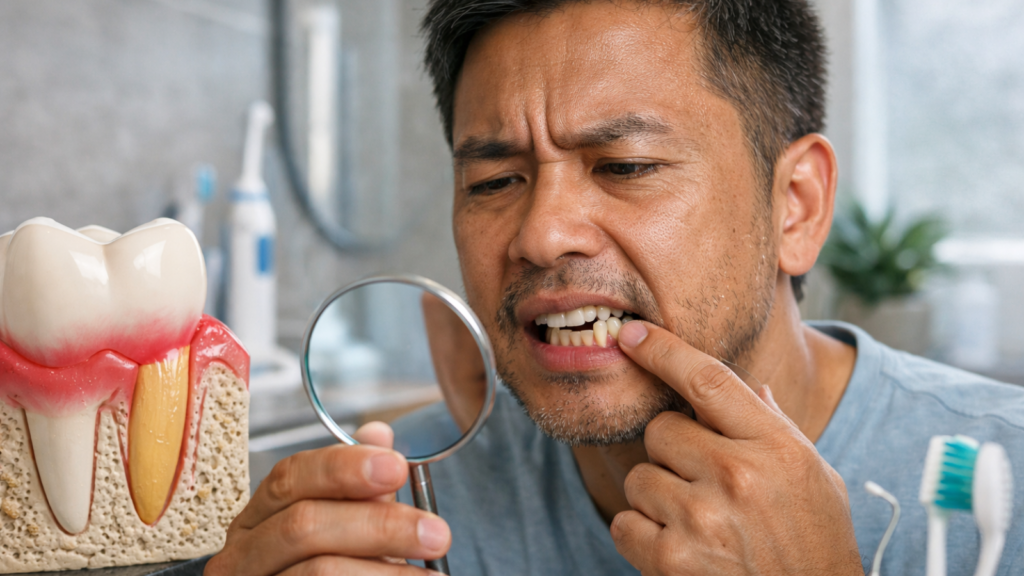
Gum recession is a condition where the gum tissue surrounding the teeth gradually wears away or pulls back, exposing more of the tooth or the tooth’s root. This progressive change affects millions of people worldwide and represents one of the most common dental concerns we see at dental clinics in Kathmandu. The exposed root surface creates both aesthetic and functional challenges that range from mild sensitivity to serious structural problems.
Understanding gum recession matters because early recognition allows for intervention before permanent damage occurs. The condition progresses slowly in most cases, giving patients and dentists time to implement protective measures. This guide explains the mechanisms behind gum recession, clarifies what can and cannot reverse naturally, and outlines the treatment pathways available in Kathmandu’s dental landscape.
Gum Recession Basics: What It Is and Why It Happens
Gum recession happens when the gumline pulls back and exposes more of the tooth or root, often starting so slowly that people don’t notice it early. It may come from gum inflammation, brushing trauma, or naturally thin gums, and it can affect both comfort and smile appearance. While gums usually don’t fully “grow back” on their own, recession can often be stabilized and managed with the right care.
What is gum recession (receding gums)?
Gum recession occurs when the margin of gum tissue that surrounds the teeth wears away or pulls back, revealing more of the tooth structure or the root surface beneath. The healthy gum attachment sits snugly around each tooth at a specific level. Recession disrupts this seal and exposes areas never meant to face the oral environment.
The root surface differs fundamentally from the crown portion of the tooth. Enamel protects the visible crown with a hard, mineralized shield. Roots lack this protective layer and instead have cementum, a much softer tissue vulnerable to decay and mechanical wear. Exposure shifts the entire tooth ecosystem toward instability.
Recession typically measures in millimeters. A 2-millimeter loss might sound minor, but it represents significant structural compromise. The attachment apparatus, composed of gum tissue, periodontal ligament, and supporting bone, loses its grip incrementally. Each millimeter of recession reduces the foundation that anchors the tooth.
Is gum recession common, and is it always gum disease?
Gum recession affects approximately 88% of people over age 65 and about 50% of people aged 18 to 64 in at least one site, making it extremely common. These prevalence figures come from large population studies and show that recession increases steadily with age. Nearly everyone experiences some degree of recession if they live long enough.
Gum disease (gingivitis and periodontitis) causes the majority of recession cases, but recession develops through multiple pathways. Aggressive brushing damages healthy gums in younger patients who maintain excellent plaque control. Thin gum tissue predisposes certain individuals to recession regardless of hygiene practices. Orthodontic movement occasionally pushes roots beyond the protective bone and gum envelope.
The distinction matters for treatment planning. Inflammation-driven recession requires infection control before any rebuilding can succeed. Mechanical recession from brushing trauma needs technique correction and protective restoration. Anatomical recession in thin tissue may benefit from grafting even in the absence of disease. Misidentifying the cause leads to ineffective treatment approaches.
Can gums grow back naturally, or is the change permanent?
Gum tissue does not regenerate or grow back to its original position once recession has occurred; the structural change remains permanent without surgical intervention. This biological reality frustrates many patients who hope that improved brushing alone will restore lost tissue. The attachment apparatus operates under strict healing limitations that prevent spontaneous regrowth.
Minor inflammation-related swelling can create the illusion of recession reversal. Swollen, puffy gums sit higher on the tooth and mask the true attachment level. These gums resolve when inflammation subsides, revealing the actual recession that existed all along. The improvement reflects reduced swelling rather than tissue regeneration.
Stabilization represents the realistic goal for non-surgical approaches. Stopping further recession preserves the remaining attachment and prevents progression. Studies show that eliminating causative factors, plaque, trauma, bite stress, halts recession in approximately 70 to 80% of cases. The existing recession remains, but the condition becomes dormant rather than active.
Surgical grafting procedures offer the only method to cover exposed roots and restore tissue height. Success rates for coverage vary from 60% to 95% depending on recession classification, surgical technique, and patient factors. Complete root coverage occurs most reliably in shallow, narrow recession defects without bone loss.
Gum recession vs periodontitis: how they’re related but not the same
Recession describes the position of the gum margin, while periodontitis defines an inflammatory disease process that destroys the supporting structures of teeth, including bone and attachment fibers. The two conditions overlap frequently but remain distinct entities with different diagnostic criteria and treatment priorities.
Periodontitis always produces recession as the disease advances. Bacterial infection triggers an inflammatory cascade that destroys collagen fibers and resorbs supporting bone. The gum margin follows the bone level downward, creating recession alongside deepening pockets. Attachment loss progresses apically (toward the root tip) from multiple directions simultaneously.
Recession can exist without periodontitis in several scenarios. Thin tissue biotype, aggressive brushing, and anatomical factors produce recession without pocket formation or bone loss. These cases show gum migration with intact attachment on the remaining tissue. Clinical examination reveals no bleeding, minimal plaque, and healthy tissue color despite the recession.
The treatment divergence becomes critical at this junction. Periodontitis demands infection control through deep cleaning (scaling and root planing), sometimes antibiotics, and strict maintenance protocols. Recession without periodontitis requires gentler interventions: technique modification, desensitizing treatments, and protective restorations. Applying periodontal therapy to mechanical recession wastes resources and misses the actual problem.
Why Gums Pull Back: Causes and Risk Factors You Should Know
The most common reason gums recede is long-term plaque buildup that inflames the gums and weakens the support around teeth. Brushing too hard, using a hard-bristle brush, grinding/clenching, and misaligned teeth can speed up gumline wear and stress. Local irritants like overhanging fillings or poorly fitting crowns can trap plaque and make recession worse in one specific area.
Plaque, gingivitis, and periodontitis: the most common pathway
Bacterial plaque accumulation initiates an inflammatory response that, left unchecked, progresses from reversible gingivitis to destructive periodontitis with accompanying recession. This infection-driven pathway accounts for roughly 60 to 70% of recession cases globally and represents the primary modifiable risk factor for most patients.
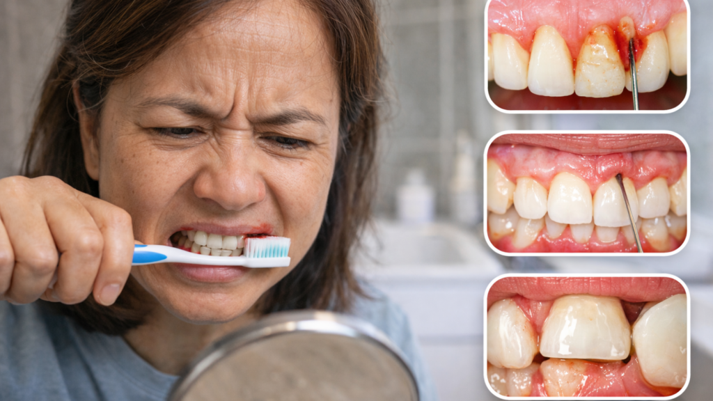
- Plaque biofilm forms continuously on tooth surfaces through bacterial colonization. The film matures over 24 to 48 hours, developing more pathogenic species as it ages. These bacteria release toxins and enzymes that trigger immune responses in the surrounding gum tissue. Early inflammation produces swelling, redness, and bleeding, the classic signs of gingivitis.
- Gingivitis remains reversible through plaque removal and improved hygiene. The inflamed tissue heals completely within 7 to 14 days when plaque control improves. Chronic gingivitis, however, transitions to periodontitis in susceptible individuals. Genetic factors, smoking, diabetes, and stress influence who progresses and how quickly.
- Periodontitis destroys the attachment apparatus through several mechanisms. Inflammatory mediators activate osteoclasts that resorb bone. Collagenases break down periodontal ligament fibers. The gum margin migrates apically to follow the bone level, creating recession and pocket formation simultaneously. This destruction occurs in episodic bursts rather than continuous, linear progression.
Brushing too hard or wrong: technique, toothbrush type, and abrasions
Excessive brushing force, horizontal scrubbing motions, and stiff-bristled toothbrushes mechanically traumatize gum tissue and create V-shaped notches at the gumline called abrasion lesions. This toothbrush trauma ranks as the second most common cause of recession in populations with good overall hygiene practices.
The mechanical damage follows predictable patterns. Prominent canines and premolars recede most frequently because patients apply extra force to these accessible surfaces. Right-handed individuals often create more recession on the left side of the mouth where their brushing arc generates maximum pressure. The recession typically appears narrow and triangular with sharp, defined borders.
Bristle stiffness plays a significant role in damage potential. Hard and medium bristles concentrate force over smaller surface areas, concentrating trauma. Soft and extra-soft bristles distribute the same brushing pressure across more contact points, reducing tissue injury. Studies demonstrate that soft bristles remove plaque equally effectively while causing 45% less gingival abrasion over 12-month periods.
Horizontal scrubbing motions concentrate force at the gumline where the brush reverses direction. Each stroke pushes tissue downward against the tooth, gradually separating the gum attachment. The proper modified Bass technique angles bristles at 45 degrees into the gumline with gentle circular or vibrating motions. This method cleans the sulcus without traumatizing the tissue margin.
Thin gums and genetics (gum biotype): why some people are more prone
Gum tissue thickness, known as gingival biotype, varies genetically among individuals, with thin biotypes (less than 1 millimeter thick) showing three to four times higher recession risk compared to thick biotypes. This anatomical predisposition explains why some patients develop recession despite excellent hygiene while others maintain stable gums despite mediocre care.
Biotype assessment occurs during routine examination. Dentists probe gently to feel tissue thickness and observe color and texture characteristics. Thin biotypes appear delicate, translucent, and pale, allowing visibility of the underlying tooth and bone. Thick biotypes feel firm, display opaque coral-pink color, and resist minor trauma without immediate change.
The structural vulnerability of thin tissue stems from reduced collagen density and narrower vascular supply. Thin gums contain fewer blood vessels per unit volume, compromising their healing response and resistance to inflammation. The reduced collagen framework provides less mechanical strength against brushing forces and bacterial invasion.
Ethnic and familial patterns emerge in biotype distribution. South Asian populations typically show intermediate biotype prevalence between European (more thick biotypes) and East Asian (more thin biotypes) patterns. Kathmandu dental patients commonly present with moderate-to-thin biotypes, making recession a particular concern in our local practice context.
Bite stress, clenching/grinding, and “abfraction”: can force worsen recession?
Excessive occlusal forces from grinding (bruxism), clenching, and malocclusion can contribute to recession by creating flexure stress at the tooth’s cervical area, though the direct causative relationship remains debated in dental research. The mechanical loading hypothesis suggests that bite forces flex teeth microscopically, fracturing enamel bonds at the gumline and predisposing those areas to both abfraction lesions and accompanying recession.
Abfraction lesions appear as wedge-shaped notches at the gumline, distinct from abrasion’s broader V-shape. These notches form on the tension side of a flexing tooth, typically the facial surface where the tooth bends outward under load. The enamel microfractures in this high-stress zone, and subsequent toothbrush abrasion or acidic erosion removes the weakened structure.
Clinical observation shows strong correlation between abfraction lesions and concurrent recession. Patients with heavy wear facets on their biting surfaces frequently display cervical notching and gum migration. The lesions appear most commonly on premolars and canines, teeth that bear significant lateral forces during chewing and grinding. If you suspect teeth grinding (bruxism) is contributing to your recession, understanding the signs and damage patterns is an important first step.
The controversy centers on whether occlusal forces directly cause recession or simply accelerate damage in the presence of other factors. Research demonstrates that force alone rarely produces recession in healthy, thick tissue with good plaque control. The combination of force plus plaque, or force plus thin tissue, or force plus trauma creates exponentially higher recession risk than any single factor.
Dental work and local irritants: overhanging fillings, crowns, and plaque traps
Improperly contoured dental restorations create plaque traps and irritation points that promote localized recession, particularly when filling margins extend beneath the gumline or crown margins compress tissue. These iatrogenic (treatment-related) factors rank among the most preventable causes of recession, yet they commonly appear in mouths with extensive restorative work.
Overhanging filling margins trap plaque in areas inaccessible to brushing and flossing. The bacterial accumulation triggers chronic inflammation specifically adjacent to the overhang. The gum tissue swells initially but eventually recedes as the chronic inflammation persists. Composite resins and amalgam fillings both create overhangs when placement technique allows excess material to extend beyond the preparation margin.
Subgingival crown margins place the restoration edge beneath the gum tissue for aesthetic purposes. Proper execution requires delicate technique to avoid violating the biological width, the 2 to 3 millimeters of space required for healthy gum attachment. Crowns that invade this space provoke chronic inflammation and recession as the body attempts to re-establish normal biological dimensions.
Orthodontic appliances create temporary plaque traps during active treatment. Brackets, wires, and elastics complicate hygiene and increase plaque accumulation around tooth necks. Most orthodontic recession develops during treatment and stabilizes after appliance removal when hygiene improves. Thin tissue biotypes show higher recession rates during orthodontic movement compared to thick biotypes.
How Dentists Diagnose and “Stage” Gum Recession
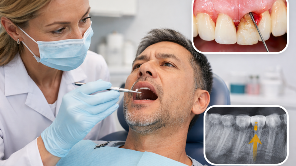
Dentists diagnose gum recession by measuring gum levels and checking pocket depth and attachment to see whether gum disease is also present. They may use X-rays to look for bone loss and to decide if the problem is mainly recession, periodontitis, or both together. Simple staging helps predict whether root coverage is realistic and whether you should see a periodontist for advanced options.
What your dentist measures: gum levels, pockets, and attachment loss
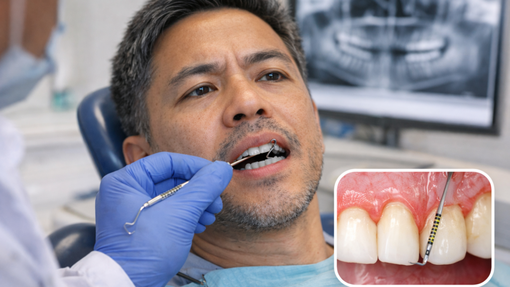
Recession assessment requires three key measurements at each tooth: the gingival margin position relative to the cementoenamel junction (CEJ), probing depth, and clinical attachment level. These measurements create a three-dimensional map of tissue architecture that guides treatment planning and monitors changes over time.
The cementoenamel junction serves as the anatomical reference point where the enamel crown meets the root cementum. This junction remains stable throughout life unless damaged by decay or abrasion. Measuring from the CEJ to the gum margin quantifies recession in millimeters. A 3-millimeter distance indicates 3 millimeters of root exposure.
Probing depth measures from the gum margin to the bottom of the gingival sulcus (the space between tooth and gum). A periodontal probe slides gently into this space using controlled pressure (approximately 25 grams). Healthy sulci measure 1 to 3 millimeters deep. Depths exceeding 4 millimeters indicate pocket formation from attachment loss or gum swelling.
Clinical attachment level combines recession and pocket depth to calculate total attachment loss. The formula adds recession (CEJ to margin) plus pocket depth (margin to sulcus base) to determine how far the attachment has migrated from its original position. This combined measurement reveals true periodontal destruction better than either measurement alone.
Do you need X-rays or scans? When imaging matters
Dental X-rays become necessary when recession accompanies periodontal pockets, bone loss, or when planning surgical grafting procedures, but mild recession without these complications often requires no imaging. The decision to radiograph balances diagnostic benefit against radiation exposure and cost considerations.
Periapical X-rays capture individual tooth roots and surrounding bone with high detail. These images reveal bone levels, root anatomy, and hidden decay on exposed root surfaces. Dentists typically order periapicals when examining recession on specific problem teeth or when planning grafting surgery to assess bone support and root morphology.
Panoramic X-rays provide a full-mouth overview showing all teeth, jaws, and sinuses in a single image. This wide view helps assess overall bone levels and identifies generalized bone loss patterns characteristic of periodontitis. The resolution remains lower than periapicals, making panoramic films better for screening than detailed diagnosis.
Cone beam computed tomography (CBCT) scans generate three-dimensional bone and tooth structure images. This advanced imaging helps plan complex grafting cases where bone thickness and root proximity need precise measurement. The radiation dose and cost (typically NPR 3,000 to NPR 8,000 in Kathmandu) limit CBCT to cases where two-dimensional X-rays provide insufficient information.
Stages/classes of recession (explained simply) and what it means for outcomes
The Miller Classification system categorizes recession into four classes based on severity and predictability of root coverage, with Class I and II showing better treatment outcomes (80 to 95% coverage) than Class III and IV (50 to 70% or less coverage). This staging system helps dentists set realistic expectations and choose appropriate treatment approaches.
- Class I recession extends into the movable gum tissue but does not reach the mucogingival junction (the line between attached gum and loose cheek tissue). No bone loss or tooth malpositioning exists. These defects respond excellently to grafting procedures because adequate bone support remains. Coverage success exceeds 90% in most published studies.
- Class II recession extends beyond the mucogingival junction into the loose tissue but maintains intact interdental bone and gum levels. The recession affects only the facial surface while the bone between teeth remains healthy. Grafting achieves 85 to 95% root coverage in these cases, though the interdental papilla (gum triangle between teeth) may not fully regenerate.
- Class III recession extends beyond the mucogingival junction with accompanying loss of interdental bone or tissue. The bone resorption or tooth malposition prevents complete root coverage even with grafting. Surgeons target partial coverage (60 to 80%) to improve aesthetics and reduce sensitivity rather than pursuing complete coverage.
- Class IV recession involves severe interproximal bone loss that eliminates the possibility of significant root coverage. Treatment focuses on halting progression, managing sensitivity, and sometimes extracting the tooth for replacement. Grafting rarely succeeds in these advanced cases because insufficient bone support remains to anchor new tissue.
When to see a periodontist instead of only a general dentist
Referral to a periodontist becomes appropriate when recession involves Class III or IV defects, accompanies active periodontitis with pockets exceeding 5 millimeters, requires complex grafting techniques, or fails to respond to general dentist treatment. General dentists manage straightforward recession cases effectively, but specialized training offers advantages in complex situations.
Periodontists complete three additional years of training after dental school focused exclusively on gum disease and supporting structures. This specialization includes advanced surgical techniques for grafting, regeneration, and implant placement. The depth of experience managing complicated recession patterns and performing microsurgical procedures exceeds typical general practice exposure.
Active periodontal disease with systemic complications, poorly controlled diabetes, smoking, immunosuppression, benefits from specialist involvement. These patients require aggressive infection control, possible antibiotic therapy, and close monitoring to prevent tooth loss. The treatment intensity and complexity justify the additional specialist consultation fee (typically NPR 1,000 to NPR 2,000 in Kathmandu).
Multiple sites requiring grafting, thin tissue biotypes with aesthetic concerns, or previous graft failures typically necessitate specialist care. The surgical complexity and precision required for optimal outcomes increase significantly in these scenarios. Most general dentists recognize these limitations and maintain referral relationships with trusted periodontists.
Symptoms and Complications: What Recession Can Lead To
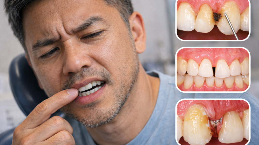
Early gum recession often shows up as cold sensitivity, teeth looking longer, or small notches near the gumline that catch your fingernail. As more root becomes exposed, the risk of root cavities increases because root surfaces are softer than enamel and decay faster. In more serious cases, you may notice black triangles, persistent bad breath, bleeding, or even tooth looseness if gum disease is involved.
Early signs: sensitivity, longer-looking teeth, and gumline notches
The first noticeable symptoms of recession typically include cold sensitivity when drinking or eating, a visual perception that teeth appear longer than before, and development of small notches or grooves at the gumline. These early warning signs often develop gradually over months to years, making them easy to dismiss until the changes become pronounced.
Root sensitivity occurs when exposed cementum and dentin allow thermal and osmotic stimuli to reach nerve endings. The exposed root surface contains thousands of microscopic tubules that connect directly to the nerve chamber. Cold liquids, sweet foods, and air exposure trigger sharp, brief pain that subsides quickly once the stimulus removes.
The “long tooth” appearance reflects actual root exposure rather than tooth growth. Adult teeth do not elongate after eruption. The recession creates an optical illusion of length by exposing root structure normally hidden beneath gums. Patients often notice this change when comparing current photographs to older images or when family members comment on smile changes.
Gumline notches develop from the combined effects of flexure stress, abrasion, and erosion acting on exposed root surfaces. These wedge-shaped defects collect plaque and stain easily. The notches feel rough to the tongue and create retention areas that complicate hygiene efforts. Sharp notch edges can irritate lip and cheek tissue during eating or speaking.
Root decay risk: why exposed roots can get cavities faster
Exposed root surfaces develop cavities three to four times more rapidly than enamel-covered crowns because cementum and dentin contain significantly less mineral content and greater porosity. Understanding how cavities form and why root surfaces are especially vulnerable helps explain this increased risk, particularly in older patients with multiple recession sites and reduced saliva flow.
Enamel consists of approximately 96% inorganic mineral crystals arranged in a dense, impermeable structure. This composition makes enamel the hardest substance in the human body and highly resistant to acid dissolution. Root surfaces contain only 65% mineral content with the remainder composed of organic collagen matrix and water.
The critical pH for enamel demineralization hovers around 5.5, requiring fairly acidic conditions before dissolution begins. Root surfaces demineralize at pH 6.0 to 6.5, levels commonly reached during normal eating and drinking. Even mildly acidic foods like fruit, coffee, and bread can initiate root surface decay in susceptible individuals.
Saliva flow becomes critically important in protecting exposed roots. Adequate saliva bathes the teeth in calcium, phosphate, and fluoride ions that remineralize early decay. Saliva also buffers acids and washes away food debris. Patients with dry mouth from medications, aging, or medical conditions face exponentially higher root decay risk when recession exposes vulnerable surfaces.
Aesthetic changes: black triangles, uneven gumline, and smile shifts
Recession creates visible aesthetic problems including dark triangular spaces between teeth (black triangles), irregular gumline contours that disrupt smile symmetry, and apparent tooth lengthening that ages the facial appearance. These cosmetic concerns often motivate patients to seek treatment even when functional symptoms remain minimal.
Black triangles form when the interdental papilla (gum tissue that fills the space between teeth) fails to reach the contact point where adjacent teeth touch. The empty triangular void appears dark against the oral cavity background. Recession eliminates the bone and tissue foundation supporting this papilla, causing it to shrink downward and exposing the gap.
Uneven gumlines create asymmetric smiles that draw attention during conversation and photographs. The gum margin normally follows a gentle scalloped pattern with peaks on canines and central incisors. Recession disrupts this harmony by creating irregular peaks and valleys. One or two receded teeth in the smile zone can dominate the overall aesthetic impression.
The aged appearance stems from the natural association between recession and advanced age. Studies show that people subconsciously estimate age partially from tooth length visibility. Extensive recession adds perceived years to facial age assessments. Younger patients with recession often report feeling self-conscious about looking older than their actual age. For patients where aesthetics are a priority alongside gum health, exploring a smile makeover consultation can help map out a combined cosmetic and periodontal treatment plan.
Red flags: pain, pus, loose teeth, or rapid worsening; when it’s urgent
Immediate dental evaluation becomes necessary when recession accompanies severe symptoms such as spontaneous pain, abscess formation with pus drainage, increasing tooth mobility, or rapid recession progression over weeks rather than months. These warning signs indicate active disease processes that can cause permanent damage if left untreated.
Spontaneous pain, discomfort that occurs without external triggers, suggests infection has reached the tooth nerve or periodontal abscess has formed. This differs from sensitivity triggered by cold or pressure. The pain may throb, persist for hours, and intensify at night. Abscesses require drainage and often antibiotics to prevent spread to deeper facial spaces.
Pus drainage appears as white or yellow fluid oozing from the gumline, often accompanied by bad taste and odor. This purulent discharge indicates active bacterial infection within a periodontal pocket. The infection destroys bone and attachment rapidly, often causing several millimeters of additional recession in just weeks without treatment.
Tooth mobility beyond normal physiological movement signals advanced attachment loss. Healthy teeth display slight movement (approximately 0.1 to 0.2 millimeters) during normal function. Recession accompanied by visible looseness or the ability to move teeth with tongue pressure indicates critical bone loss requiring urgent intervention to prevent tooth loss.
Rapid progression, noticeable recession changes within 4 to 8 weeks, suggests aggressive periodontitis or acute trauma. Normal recession advances slowly over years. Accelerated progression requires immediate diagnosis to identify and eliminate causative factors. Delayed treatment often results in tooth loss that might have been prevented with timely intervention.
Can Gum Recession Be Reversed? What Actually Works at Home
Most people can’t truly reverse recession at home, but they can often stop it from progressing and reduce sensitivity with correct daily habits. Gentle brushing with a soft brush, proper flossing/interdental cleaning, and fluoride-based products help protect exposed roots and calm inflammation. Avoid abrasive powders, harsh whitening, and “gum regrowth” claims, because they can irritate tissues and worsen recession over time.
“Reversing” vs “stopping”: what realistic improvement looks like
Home care cannot reverse existing recession or regenerate lost gum tissue, but proper techniques can halt progression in 70 to 85% of cases and occasionally produce 0.5 to 1.0 millimeter of apparent improvement through inflammation reduction and tissue rebound. Setting realistic expectations prevents disappointment and helps patients appreciate the significant value of stabilization.
True reversal requires surgical intervention to physically reposition tissue or graft new material over exposed roots. No amount of improved brushing, special toothpastes, or natural remedies will cause tissue migration back up the root surface. The attachment apparatus lacks the cellular mechanisms needed for spontaneous upward migration once recession has occurred.
Inflammation reduction creates minor visual improvement that patients sometimes interpret as reversal. Swollen, inflamed gums sit higher on teeth than healthy tissue. Removing plaque and improving hygiene resolves this swelling over 2 to 3 weeks, causing the gum margin to drop slightly and reveal the true recession extent. The tissue appears healthier and firmer despite sitting lower than before treatment.
Tissue rebound describes a small upward migration (typically 0.5 to 1.0 millimeter) that sometimes follows elimination of chronic irritation. This minor improvement likely results from collagen fiber reorganization and resolution of chronic edema rather than true attachment gain. Studies document this rebound in approximately 30 to 40% of treated recession sites, though the effect remains modest.
The best brushing method for recession (gentle technique that protects gums)
The modified Bass technique, using soft or extra-soft bristles at a 45-degree angle to the gumline with gentle circular or vibrating motions, provides optimal plaque removal while minimizing trauma to receded or vulnerable gum tissue. This method cleans effectively without the destructive horizontal scrubbing that causes mechanical recession.
Brush selection forms the foundation of safe technique. Soft and extra-soft bristles (0.15 to 0.18 millimeter diameter) bend easily against tissue, distributing pressure over larger surface areas. Medium and hard bristles (0.20 to 0.25 millimeter diameter) concentrate force at their tips and resist bending, increasing trauma potential. Electric brushes with pressure sensors help prevent excessive force.
The 45-degree angle positions bristle tips into the gingival sulcus where plaque accumulates most heavily. The bristles should contact both the tooth surface and gum margin simultaneously. Gentle pressure, just enough to blanch the tissue white, allows bristles to penetrate the sulcus without traumatizing attachment fibers.
Motion pattern determines whether cleaning damages or protects tissue. Small circular or short back-and-forth vibrating motions dislodge plaque without pushing the gum margin downward. Each tooth requires 10 to 15 seconds of focused cleaning time. Rushing through brushing invariably leads to increased pressure and horizontal scrubbing as patients try to compensate for insufficient time with additional force.
Flossing and interdental cleaning: what to use if gaps or black triangles exist
Traditional floss works effectively in tight contacts without recession, but patients with recession, black triangles, or wider interdental spaces achieve better results using interdental brushes, soft picks, or water flossers that clean exposed root surfaces and open embrasure spaces. The tool selection must match the anatomy created by recession.
Interdental brushes consist of small bristled tips on wire handles sized to fit between teeth. These brushes come in diameter ranges from 0.4 millimeters (pink, finest) to 1.5 millimeters (black, largest). Proper sizing allows the brush to slide between teeth with slight resistance but without forcing. The bristles contact both adjacent tooth surfaces and the gum papilla, removing plaque effectively from all surfaces.
Soft picks combine flexibility with cleaning efficiency in moderately receded areas. The rubber tips compress during insertion and expand once through the contact point, conforming to irregular embrasure shapes. Studies show soft picks remove 78 to 85% as much plaque as interdental brushes while causing less trauma in sensitive areas.
Water flossers direct pulsating water streams into interdental spaces and beneath the gumline. This irrigation removes loose debris and bacteria but shows less effectiveness than mechanical cleaning for disrupting mature plaque biofilm. The combination of interdental brush followed by water flossing provides superior cleaning compared to either method alone.
Sensitivity relief: fluoride, desensitizing toothpaste, and protecting exposed roots
Exposed root sensitivity improves through three main approaches: high-concentration fluoride applications that occlude dentinal tubules, desensitizing toothpastes containing potassium nitrate or stannous fluoride used twice daily, and physical barriers such as fluoride varnish or bonding agents applied by dentists. Most patients require multiple complementary approaches for optimal relief.
High-concentration fluoride (5,000 parts per million prescription toothpaste or 22,600 parts per million varnish) precipitates calcium fluoride crystals within dentinal tubules. These crystals physically block the fluid flow that triggers nerve responses. The effect builds gradually over 2 to 4 weeks of consistent application. Prescription fluoride toothpastes cost NPR 1,200 to NPR 2,000 in Kathmandu pharmacies.
Desensitizing toothpastes work through two mechanisms depending on active ingredients. Potassium nitrate formulations (8% concentration) depolarize nerve fibers, reducing their excitability and pain response. Stannous fluoride and calcium sodium phosphosilicate (NovaMin) physically occlude tubules similar to high-concentration fluoride. Maximum benefit requires 4 to 6 weeks of twice-daily use.
Professional fluoride varnish application provides immediate but temporary relief lasting 3 to 6 months. Dentists paint the concentrated fluoride resin onto dried, receded roots where it hardens on contact with saliva. The varnish slowly releases fluoride while physically sealing exposed tubules. Reapplication every 3 to 4 months maintains protection in severely affected areas.
Bonding agents and resin restorations permanently seal exposed roots when sensitivity persists despite other measures. Dentists etch the root surface and apply adhesive resin that bonds chemically to the tooth structure. This approach works particularly well for abfraction notches where the concave defect retains the bonding material reliably.
Myth-busting: salt, oils, powders, and “gum regrowth” claims; what to avoid
Popular home remedies including salt rinses, oil pulling, charcoal powders, and herbal supplements lack scientific evidence for reversing recession and may cause harm through abrasive damage or delayed professional treatment of active disease. Understanding why these approaches fail helps patients avoid wasted time and resources.
Salt water rinses provide temporary anti-inflammatory effects through osmotic action but cannot prevent or reverse recession. The high salt concentration draws fluid from swollen tissue, reducing edema temporarily. This effect lasts only hours and does nothing to address underlying causes such as plaque accumulation or brushing trauma. Patients often interpret temporary swelling reduction as healing when the recession remains unchanged.
Oil pulling involves swishing coconut, sesame, or sunflower oil for 10 to 20 minutes daily. Proponents claim this traditional Ayurvedic practice removes toxins and promotes gum regeneration. Systematic reviews of published studies find no evidence supporting recession reversal or tissue regeneration. The practice may reduce plaque similarly to any prolonged swishing with any liquid, but offers no advantages over standard brushing and flossing.
Activated charcoal powders and toothpastes have gained popularity for teeth whitening and oral health. The charcoal’s abrasive particles scratch enamel and can accelerate wear on already-exposed roots. No evidence supports gum regeneration claims. The American Dental Association (ADA) has not approved any charcoal products due to insufficient safety and efficacy data.
Herbal supplements marketed for gum health, turmeric, coenzyme Q10, green tea extract, show anti-inflammatory properties in laboratory studies but lack clinical evidence for preventing or reversing recession in humans. These products should not replace evidence-based treatment. Patients who delay professional care while pursuing alternative approaches risk progression to irreversible attachment loss.
Treatment Options: From Stabilizing Recession to Covering Exposed Roots
Professional treatment depends on the cause and severity, ranging from cleaning and deep cleaning to gum grafting or minimally invasive procedures to cover exposed roots. If there are gumline notches or root defects, a combined plan like bonding plus gum treatment may provide better comfort and durability. Aftercare matters as much as the procedure, so healing instructions, maintenance cleanings, and technique corrections are key to preventing recurrence.
Professional cleaning and deep cleaning: when it’s enough (and when it isn’t)
Standard professional cleaning (scaling and polishing) suffices for mild recession without periodontitis, while deep cleaning (scaling and root planing) becomes necessary when recession accompanies pockets measuring 4 millimeters or deeper with subgingival calculus and active inflammation. The distinction determines both treatment invasiveness and cost expectations.
Standard scaling removes supragingival (above the gumline) plaque and calculus using ultrasonic instruments and hand scalers. This preventive procedure typically requires 30 to 45 minutes. Our professional teeth cleaning service covers this baseline care, which costs NPR 2,000 to NPR 3,000 in Kathmandu dental clinics. Scaling alone stabilizes recession caused by mild plaque accumulation and poor hygiene when no periodontal disease exists.
Deep cleaning treats periodontal disease by removing subgingival (below the gumline) calculus and diseased cementum from root surfaces. The procedure often requires local anesthesia and extends over multiple appointments to thoroughly clean all affected areas. Costs range from NPR 8,000 to NPR 15,000 for full-mouth treatment depending on disease severity and clinic location.
Root planing smooths the root surface after calculus removal, creating an environment less conducive to bacterial recolonization. Rough root surfaces harbor bacteria in microscopic irregularities that brushing cannot reach. The smoothing process removes these retention areas and allows gum tissue to reattach more closely to the cleaned root surface.
Deep cleaning alone cannot reverse existing recession or provide root coverage. The procedure halts disease progression and creates conditions favorable for tissue stabilization. Studies show that 65 to 75% of patients maintain stable recession levels for 3 to 5 years following successful deep cleaning and compliance with maintenance visits every 3 to 4 months.
Gum graft surgery types (CT graft, free gingival graft, substitutes) and who needs what
Three primary grafting approaches exist: connective tissue grafts (most common, 85 to 95% root coverage success), free gingival grafts (builds thick tissue, 70 to 80% coverage), and acellular dermal matrix substitutes (avoids palate harvesting, 75 to 85% coverage). The choice depends on recession classification, aesthetic importance, tissue availability, and patient preferences regarding donor site surgery.
Connective tissue grafts harvest tissue from beneath the palate surface, leaving the outer layer intact. The surgeon creates a small flap on the palate roof, removes the subepithelial connective tissue layer, and closes the flap with sutures. This technique provides adequate graft thickness for root coverage while healing the donor site by primary intention (edges directly reattached) with minimal discomfort.
The harvested connective tissue positions over the receded root and secures with sutures to adjacent gum tissue. The overlying gum flap repositions coronally (toward the crown) to cover the graft and exposed root. Final color and texture match surrounding tissue well because the graft integrates beneath the patient’s own tissue. Healing requires 2 to 3 weeks for initial tissue integration and 3 to 6 months for complete maturation.
Free gingival grafts remove the full thickness of palatal tissue including both connective tissue and overlying epithelium. This approach creates a distinct white or lighter-colored patch of thick tissue over the recession site. Dentists use free gingival grafts primarily to create a wider band of attached tissue rather than for aesthetic root coverage. The thick tissue resists future recession better than native thin tissue.
Acellular dermal matrix (ADM) grafts use processed donor tissue from tissue banks, eliminating the need for palate surgery. The ADM provides a collagen scaffold that patient cells repopulate during healing. Success rates approximate connective tissue grafts (75 to 85% coverage) with the advantage of single-site surgery. Costs increase by NPR 15,000 to NPR 25,000 compared to connective tissue grafts due to material expenses.
Minimally invasive options (pinhole/tunneling/CAF): who may be a candidate
Minimally invasive recession coverage techniques including pinhole surgery, tunnel grafting, and coronally advanced flaps offer faster healing and less discomfort compared to traditional grafting but succeed primarily in Class I and shallow Class II recession with intact interdental tissue. Patient selection determines success more than technique choice.
Pinhole surgical technique creates a small entry point above the recession site through which the surgeon tunnels beneath the gum tissue. Specialized instruments loosen the tissue from the underlying bone, allowing the gum to slide coronally over the exposed root. Collagen strips placed through the pinhole stabilize the repositioned tissue while healing occurs.
The pinhole approach eliminates sutures and incision lines, reducing post-operative pain and accelerating recovery. Patients typically resume normal eating within 24 to 48 hours compared to 7 to 10 days with traditional grafting. The technique works best for treating multiple adjacent recession sites simultaneously because the tunnel extends across several teeth through limited access points.
Tunnel grafting combines minimally invasive access with connective tissue reinforcement. The surgeon creates a partial-thickness tunnel extending from one recession site to adjacent areas. Connective tissue grafts slide through the tunnel to cover multiple roots through limited incisions. This hybrid approach increases predictability compared to pinhole alone while maintaining reduced invasiveness.
Coronally advanced flaps (CAF) reposition existing gum tissue coronally without adding graft material. The technique succeeds only when adequate tissue thickness and width exist to stretch over the exposed root. The surgeon creates releasing incisions, elevates the tissue, and sutures it in the coronal position. Success rates reach 85 to 90% in carefully selected Class I recession but drop to 50 to 60% in Class II cases.
Combined fixes: when bonding and gum procedures work better for notches/NCCLs
Deep abfraction lesions and cervical notches often require composite resin bonding in combination with gum grafting to achieve optimal functional and aesthetic results, as grafting alone cannot fill concave defects and bonding alone leaves exposed root surfaces vulnerable. The combined approach addresses both structural and soft tissue components.
Non-carious cervical lesions (NCCLs) create challenges for isolated grafting approaches. The concave defect shape prevents stable graft positioning because the tissue cannot adapt into the notch depression. Grafts placed over unfilled notches often fail to achieve complete coverage and may slump into the defect during healing, creating irregular contours.
Composite bonding fills the NCCL before gum surgery, creating a convex tooth contour that supports graft adaptation. The dentist etches the exposed root surface, applies bonding agent, and incrementally builds composite resin to restore the tooth’s natural cervical contour. This foundation allows the subsequent graft to position properly and achieve predictable coverage.
The staging sequence matters for optimal results. Bonding typically occurs first to allow tissue maturation around the restoration before grafting surgery. Some surgeons prefer bonding after grafting to avoid contaminating the surgical field with composite debris. Both sequences show similar long-term success (80 to 90% stable coverage) when executed properly.
Combined treatment costs range from NPR 45,000 to NPR 75,000 per tooth in Kathmandu depending on lesion severity and graft complexity. The investment typically exceeds isolated procedures by 30 to 40% but provides superior long-term stability and aesthetics compared to either approach alone.
Aftercare and prevention plan: healing timeline, maintenance visits, and stopping recurrence
Graft healing progresses through three phases: initial attachment (days 1 to 14), tissue maturation (weeks 2 to 12), and final remodeling (months 3 to 6), with specific care instructions and restrictions for each phase. Following the protocol precisely determines whether the surgery achieves its maximum potential or falls short due to preventable complications.
The first 14 days focus on protecting the surgical site from disruption. Patients avoid brushing the grafted area entirely, using only prescribed chlorhexidine rinses twice daily. Diet restrictions include soft foods requiring minimal chewing and avoidance of anything hard, crunchy, or requiring tearing motions. Suture removal occurs at 10 to 14 days when initial tissue integration has secured the graft position.
Weeks 2 through 12 allow careful hygiene resumption using extra-soft brushes and roll technique that avoids direct pressure on the graft. The tissue gradually thickens and strengthens during this period. Color and texture differences between grafted and native tissue remain apparent. Sensitivity often persists as nerve fibers reestablish connections through the maturing tissue.
Months 3 through 6 complete the remodeling phase as collagen fibers reorganize and vascularity normalizes. Final color matching occurs during this period, though some persistent color variation may remain with free gingival grafts. Complete integration allows return to normal hygiene practices using the gentle techniques that prevent recession recurrence.
Maintenance visits every 3 to 4 months become critical for long-term stability. These appointments allow professional cleaning to remove plaque accumulations patients inevitably miss, early detection of recession signs at new sites, and reinforcement of protective techniques. Studies show that patients who maintain regular professional care retain successful graft outcomes in 90 to 95% of sites over 5 to 10 years compared to 60 to 70% success in patients who neglect maintenance.
Gum recession treatment in Kathmandu: cost drivers, timeline, and how to choose a clinic
Gum recession treatment costs in Kathmandu range from NPR 2,000 for preventive scaling to NPR 60,000 to NPR 100,000 per tooth for advanced grafting procedures, with costs determined by recession severity, technique complexity, surgeon experience, and material choices. Patients seeking comprehensive dental in Kathmandu can find full-service clinics offering everything from initial consultation to surgical grafting under one roof. Understanding these variables helps patients budget appropriately and make informed decisions.
Basic treatment pathway costs (typical ranges as of January 2026):
- Initial consultation and examination: NPR 1,000 to NPR 2,000
- Scaling and polishing: NPR 2,000 to NPR 3,000
- Deep cleaning (scaling and root planing, full mouth): NPR 8,000 to NPR 15,000
- Connective tissue graft (single tooth): NPR 40,000 to NPR 65,000
- Free gingival graft (single tooth): NPR 35,000 to NPR 55,000
- Acellular dermal matrix graft (single tooth): NPR 60,000 to NPR 90,000
- Multiple-site grafting (3 to 4 adjacent teeth): NPR 80,000 to NPR 130,000
Treatment timelines extend over several months for most surgical cases. The sequence typically includes initial consultation and diagnosis (week 0), deep cleaning if needed (weeks 1 to 2), healing period (weeks 3 to 6), surgical grafting (week 6 to 8), initial healing (weeks 8 to 10), suture removal (week 10), and progressive healing over months 3 through 6. Total time from consultation to final result spans 6 to 8 months.
Choosing a qualified provider requires evaluating several factors beyond price alone. Firstly, verify formal training credentials, general dentists can perform basic grafting, but periodontists complete specialized training that improves outcomes in complex cases. Secondly, request before and after photographs of previous recession cases to assess aesthetic results. Thirdly, confirm sterilization protocols and infection control practices through facility tours.
BrightSmile Dental Clinic in Putalisadak offers comprehensive recession evaluation and treatment planning, with transparent pricing within Kathmandu’s typical ranges and individualized treatment protocols based on recession classification and patient goals. The clinic prioritizes gentle techniques and patient comfort while maintaining strict sterilization standards. Consultation appointments provide detailed assessment, treatment options explanation, and realistic outcome expectations before committing to any procedure.
Prevention remains more effective than treatment for gum recession. Patients who adopt gentle brushing techniques, maintain regular professional cleanings every 6 months, and address grinding or clenching habits preserve their gum architecture far more successfully than those attempting to restore lost tissue after recession has advanced. Early intervention when recession first appears offers the best chance for long-term stability and minimal treatment complexity. Schedule a comprehensive examination at BrightSmile Dental Clinic to assess your gum health and develop a personalized protection plan tailored to your specific risk factors and oral anatomy.
What causes gum recession, and is it always gum disease?
Gum recession occurs when the gum tissue pulls away from the teeth, exposing more of the root surface. It is commonly caused by gum disease, aggressive brushing, genetics, smoking, or bite stress. While gum disease is a leading cause, not all gum recession stems from infection; other mechanical or genetic factors may be involved.
Can receding gums grow back or be “reversed” naturally?
Receding gums do not grow back naturally once the tissue is lost. While the damage cannot be reversed without surgery, the progression can be stopped by eliminating causes like plaque, trauma, or grinding. Gum grafts or root-coverage procedures are typically required to restore coverage over exposed roots.
What are the earliest signs of gum recession I should watch for?
Early signs of gum recession include longer-looking teeth, uneven gumlines, cold sensitivity, or notches near the gumline. These changes often appear gradually and may signal gum wear, brushing trauma, or early disease. A dental checkup can confirm the cause and help prevent further recession.
Is gum recession an emergency and when should I seek urgent care?
Gum recession is not usually an emergency, but seek urgent care if you notice swelling, pus, fever, severe pain, or rapid changes. These symptoms may indicate infection or advanced gum disease. Prompt dental evaluation helps prevent complications and protects your teeth from further damage.
Can brushing too hard cause gum recession, and what technique is safest?
Brushing too hard can cause gum recession by damaging the gumline. Use a soft-bristled toothbrush and apply light pressure with small, circular motions angled toward the gums. Avoid aggressive scrubbing to prevent gum injury. A dentist can demonstrate proper technique if recession has already started.
What non-surgical treatments can stop gum recession from worsening?
Non-surgical treatments include professional cleaning, plaque control, bite adjustment, and fluoride for sensitivity. These methods can stop gum recession from progressing by eliminating causes and protecting exposed roots. Dentists may also smooth rough fillings or recommend desensitizing products to support oral health.
When is gum graft surgery recommended, and what results are realistic?
Gum graft surgery is recommended for progressing recession, exposed roots, or high sensitivity. It adds gum thickness and may cover roots for better protection and comfort. Realistic results depend on tissue quality, recession depth, and cause control. A periodontist can assess and plan accordingly.
What is the “pinhole” (minimally invasive) technique, and who is a good candidate?
The pinhole technique repositions gum tissue without grafts using small entry points. It is best for patients with mild-to-moderate recession and sufficient healthy tissue. Advanced cases with severe loss may still need grafting. A periodontist can determine eligibility based on gum thickness and recession type.
Does gum recession increase the risk of cavities, and how can I prevent root decay?
Gum recession increases cavity risk by exposing root surfaces, which are softer than enamel and decay faster. Prevent root decay by brushing gently with fluoride toothpaste, using desensitizing products, and keeping plaque off the roots. Regular dental checkups help catch root cavities early.
How much does gum recession treatment cost in Kathmandu, and what affects the price?
Gum recession treatment in Kathmandu ranges from low-cost cleanings to higher-cost surgeries. Prices vary by severity, number of affected teeth, treatment type (e.g., deep cleaning or grafting), and provider specialization. A full dental exam with measurements and X-rays provides the most accurate estimate.